

Development and characterization of a diamond-insulated graphitic multi electrode array realized with ion beam lithography
- PMID: 25558992
- PMCID: PMC4327033
- DOI: 10.3390/s150100515
Development and characterization of a diamond-insulated graphitic multi electrode array realized with ion beam lithography
Abstract
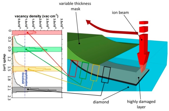
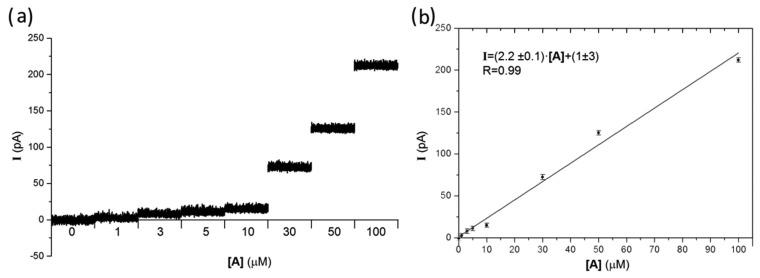
The detection of quantal exocytic events from neurons and neuroendocrine cells is a challenging task in neuroscience. One of the most promising platforms for the development of a new generation of biosensors is diamond, due to its biocompatibility, transparency and chemical inertness. Moreover, the electrical properties of diamond can be turned from a perfect insulator into a conductive material (resistivity ~mΩ·cm) by exploiting the metastable nature of this allotropic form of carbon. A 16‑channels MEA (Multi Electrode Array) suitable for cell culture growing has been fabricated by means of ion implantation. A focused 1.2 MeV He+ beam was scanned on a IIa single-crystal diamond sample (4.5 × 4.5 × 0.5 mm3) to cause highly damaged sub-superficial structures that were defined with micrometric spatial resolution. After implantation, the sample was annealed. This process provides the conversion of the sub-superficial highly damaged regions to a graphitic phase embedded in a highly insulating diamond matrix. Thanks to a three-dimensional masking technique, the endpoints of the sub-superficial channels emerge in contact with the sample surface, therefore being available as sensing electrodes. Cyclic voltammetry and amperometry measurements of solutions with increasing concentrations of adrenaline were performed to characterize the biosensor sensitivity. The reported results demonstrate that this new type of biosensor is suitable for in vitro detection of catecholamine release.
Figures


References
-
- Human Brain Project. [(accessed on 20 October 2014)]. Available online: https://www.humanbrainproject.eu/it/home.
-
- National Institute of Health The Brain Initiative. [(accessed on 20 October 2014)]. Available online: http://www.braininitiative.nih.gov/index.htm.
-
- Coupland R.E. The Natural History of the Chromaffin Cell. Longmans Green and Co Ltd.; London, UK: 1965.
-
- Wightman R.M., Jankowski J.A., Kennedy R.T., Kawagoe D.T., Schroeder T.J., Leszczyszyn D.J., Near J.A., Diliberto E.J., Jr., Viveros O.H. Temporally resolved catecholamine spikes correspond to single vesicle release from individual chromaffin cells. Proc. Natl. Acad. Sci. USA. 1991;88:10754–10758. - PMC - PubMed
-
- Ciolkowski E.L., Maness K.M., Cahill P.S., Wightman R.M., Evans D.H., Fosset B., Amatore C. Disproportionation during Electro-oxidation of Catecholamines at Carbon-Fiber Microelectrodes. Anal. Chem. 1994;66:3611–3617.
Publication types
MeSH terms
Substances
LinkOut - more resources
Full Text Sources
Other Literature Sources
