. 2015 May;17(5):579-92.
doi: 10.1016/j.jcyt.2014.12.003. Epub 2015 Jan 2.

Notch ligand Delta-like 1 promotes in vivo vasculogenesis in human cord blood-derived endothelial colony forming cells

Affiliations

Notch ligand Delta-like 1 promotes in vivo vasculogenesis in human cord blood-derived endothelial colony forming cells

Hyojin Kim et al. Cytotherapy. 2015 May.

Abstract

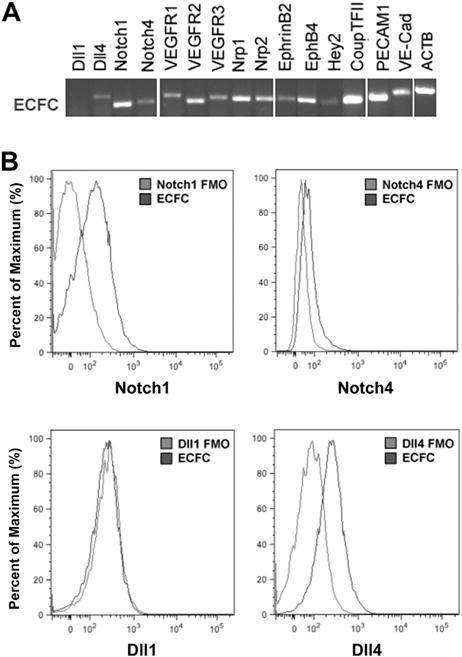
Background aims: Human cord blood (CB) is enriched in circulating endothelial colony forming cells (ECFCs) that display high proliferative potential and in vivo vessel forming ability. Because Notch signaling is critical for embryonic blood vessel formation in utero, we hypothesized that Notch pathway activation may enhance cultured ECFC vasculogenic properties in vivo.

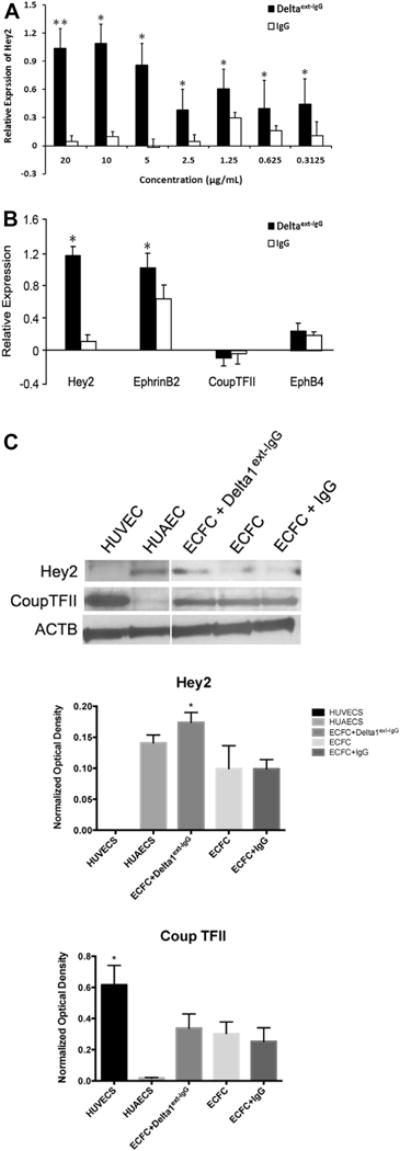
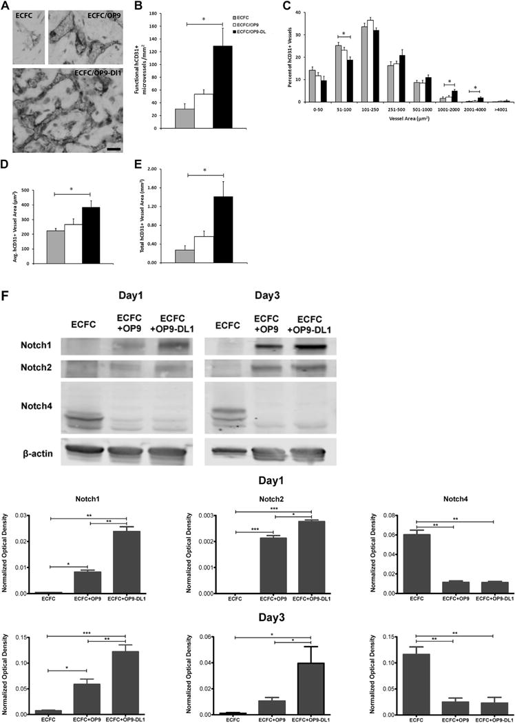
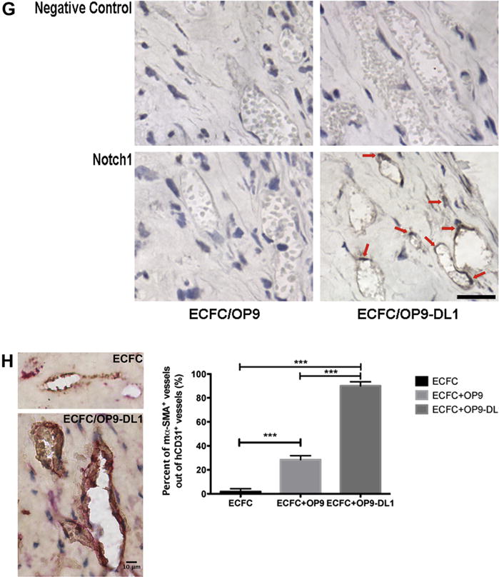
Methods: In vitro ECFC stimulation with an immobilized chimeric Notch ligand (Delta-like1(ext-IgG)) led to significant increases in the mRNA and protein levels of Notch regulated Hey2 and EphrinB2 that were blocked by treatment with γ-secretase inhibitor addition. However, Notch stimulated preconditioning in vitro failed to enhance ECFC vasculogenesis in vivo. In contrast, in vivo co-implantation of ECFCs with OP9-Delta-like 1 stromal cells that constitutively expressed the Notch ligand delta-like 1 resulted in enhanced Notch activated ECFC-derived increased vessel density and enlarged vessel area in vivo, an effect not induced by OP9 control stromal implantation.

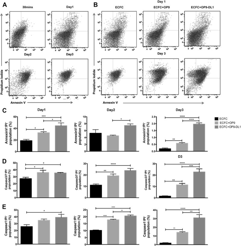
Results: This Notch activation was associated with diminished apoptosis in the exposed ECFC.

Conclusions: We conclude that Notch pathway activation in ECFC in vivo via co-implanted stromal cells expressing delta-like 1 promotes vasculogenesis and augments blood vessel formation via diminishing apoptosis of the implanted ECFC.

Keywords: Notch ligand delta-like 1 (Dll1); OP9-Delta-like 1 stromal cells (OP9-DL1); apoptosis; endothelial colony forming cells (ECFCs); vasculogenesis.


Conflict of interest statement

Disclosure of interest: The authors have no commercial, proprietary, or financial interest in the products or companies described in this article.

Figures

Figure 1
Figure 1
The expression of Notch pathway related transcripts in cultured ECFCs. (A) Human CB–derived ECFCs expressed numerous Notch pathway related transcripts and EC gene markers such as platelet endothelial cell adhesion molecule-l and VE-Cadherin. (B) Notch ligands and receptors were expressed on a subset of ECFCs. The representative histograms show comparison between fluorescence minus one (FMO) control (gray) and Notch molecule expression (black) on the surface of ECFCs.
Figure 2
Figure 2
ECFCs respond to Notch signaling with increased expression of known downstream target genes (n = 3). The dose-dependent activation of endogenous Notch signaling in human CB–derived ECFCs is indicated by Hey2 (A) expression after 3 days of incubation in various concentrations of Delta1ext-IgG. (B) Quantitative RT-PCR analysis revealed Hey2 and EphrinB2 transcripts were significantly increased after 3 days of stimulation with 10 μg/mL Delta 1ext-IgG, whereas the expression of Coup TFII and EphB4 were not significantly affected. Expression levels were presented as a fold change (in logarithmic scale) compared with baseline levels and were normalized by using ATP5B as a housekeeping gene. The expression at day 0 in nontreated cells served as a baseline value. n = 5. *P < 0.05. (C) Upper panel depicts representative immunoblots of Hey2 and Coup TFII proteins and lower panel reveals quantification (protein expression levels were normalized by using β-actin). *P < 0.05. HUAEC, human umbilical artery endothelial cells; HUVEC, human umbilical vein endothelial cells.
Figure 3
Figure 3
In vivo Dll1 stimulation boosts the formation of functional vessels (n = 3). (A) Anti-human CD31 staining identified human CB–derived ECFCs, alone or combined with OP9 or OP9-DL1, that have formed microvessels in collagen gels after 14 days of implantation. Upon the stimulation of Dll1, there was a significant increase in the number of vessels formed by human CB–derived ECFCs and perfused with murine red blood cells per mm2 in the gel (B). In addition, the size distribution of hCD31+ microvessels was noticeably altered (C), shifting toward larger-sized vessels (1001–4000 μm2) with Dll1 stimulation. Moreover, vessel morphology was significantly altered by the presence of Dll1 with increased average vessel area (D) and total vascular areas (E). (F) Upper panel shows representative immunoblots of Notch 1, Notch 2 and Notch 4 proteins and lower 2 panels show quantification of repeated experiments (protein expression levels were normalized using β-actin); n = 3. *P < 0.05. Anti-cleaved Notch 1 (Val1744) antibody staining (G) in endothelial nuclei confirms that the activation of Notch 1 is detected in vivo in newly formed human vessels in the presence of Dll1; n = 6. Scale bar represents 100 μm. (H) Anti-mouse smooth muscle α actin (αSMA) antibody staining (red) was detected in perivascular cells around human ECFC–derived CD31+ vessels (brown) in implants with OP9-DL1 stromal cells. Scale bar represents 10 μm. *P < 0.05; **P < 0.001; ***P < 0.0001.
Figure 3
Figure 3
In vivo Dll1 stimulation boosts the formation of functional vessels (n = 3). (A) Anti-human CD31 staining identified human CB–derived ECFCs, alone or combined with OP9 or OP9-DL1, that have formed microvessels in collagen gels after 14 days of implantation. Upon the stimulation of Dll1, there was a significant increase in the number of vessels formed by human CB–derived ECFCs and perfused with murine red blood cells per mm2 in the gel (B). In addition, the size distribution of hCD31+ microvessels was noticeably altered (C), shifting toward larger-sized vessels (1001–4000 μm2) with Dll1 stimulation. Moreover, vessel morphology was significantly altered by the presence of Dll1 with increased average vessel area (D) and total vascular areas (E). (F) Upper panel shows representative immunoblots of Notch 1, Notch 2 and Notch 4 proteins and lower 2 panels show quantification of repeated experiments (protein expression levels were normalized using β-actin); n = 3. *P < 0.05. Anti-cleaved Notch 1 (Val1744) antibody staining (G) in endothelial nuclei confirms that the activation of Notch 1 is detected in vivo in newly formed human vessels in the presence of Dll1; n = 6. Scale bar represents 100 μm. (H) Anti-mouse smooth muscle α actin (αSMA) antibody staining (red) was detected in perivascular cells around human ECFC–derived CD31+ vessels (brown) in implants with OP9-DL1 stromal cells. Scale bar represents 10 μm. *P < 0.05; **P < 0.001; ***P < 0.0001.
Figure 4
Figure 4
OP9-DL1 stimulation diminishes apoptosis of ECFCs in collagen gels (n = 5). (A) Representative dot plots of apoptosis analysis of ECFCs at 30 min or on days 1–3. (B) Representative dot plots of apoptosis analysis of ECFC alone, co-implanted with OP9, or OP9-DL1 on days 1–3. (C) The Annexin/PI viable population of ECFC co-implanted with OP9-DL1 was significantly higher on day 1 and 3 compared with implantation of ECFC alone or ECFC with OP9 on day 1 and 3. (D) The percentage of the Caspase 3/7/PI population of ECFC co-implanted with OP9-DL1 was significantly greater compared with ECFC alone or with OP9 on day 3. (E) ECFC co-implanted with OP9-DL1 displayed a higher percentage of Caspase 1/PI cells compared with ECFC alone or ECFC with OP9 co-implants. n = 6. *P < 0.05.

Similar articles

Cited by

References

    1. Niessen K, Karsan A. Notch signaling in cardiac development. Circ Res. 2008;102:1169–81. - PubMed
    1. Radtke F, Fasnacht N, Macdonald HR. Notch signaling in the immune system. Immunity. 2010;32:14–27. - PubMed
    1. Ables JL, Breunig JJ, Eisch AJ, Rakic P. Not(ch) just development: Notch signalling in the adult brain. Nat Rev Neurosci. 2011;12:269–83. - PMC - PubMed
    1. Swift MR, Weinstein BM. Arterial-venous specification during development. Circ Res. 2009;104:576–88. - PubMed
    1. Kume T. Novel insights into the differential functions of Notch ligands in vascular formation. J Angiogenes Res. 2009;1:8. - PMC - PubMed

Publication types

MeSH terms