

Review
doi: 10.1152/physiol.00019.2014.
In vivo fluorescence microscopy: lessons from observing cell behavior in their native environment
Affiliations
- PMID: 25559154
- PMCID: PMC4285577
- DOI: 10.1152/physiol.00019.2014
Item in Clipboard
Review
In vivo fluorescence microscopy: lessons from observing cell behavior in their native environment
Physiology (Bethesda).
2015 Jan.
Abstract
Microscopic imaging techniques to visualize cellular behaviors in their natural environment play a pivotal role in biomedical research. Here, we review how recent technical advances in intravital microscopy have enabled unprecedented access to cellular physiology in various organs of mice in normal and diseased states.
©2015 Int. Union Physiol. Sci./Am. Physiol. Soc.
Conflict of interest statement
No conflicts of interest, financial or otherwise, are declared by the author(s).
Figures
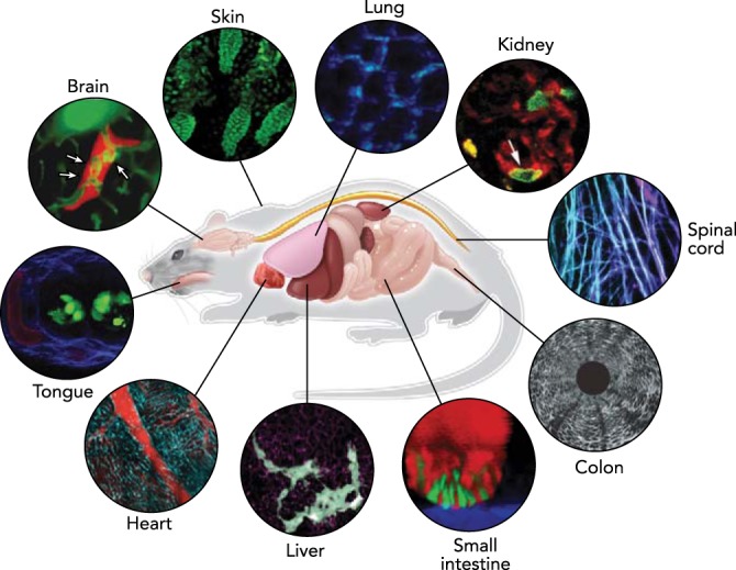
In vivo imaging in the mouse Advances in optical techniques in conjunction with sample preparation methods allow microscopic visualization of cellular dynamics in various organs in a living experimental animal. Representative images in each organ acquired by intravital microscopy on mouse models are shown. Images reproduced with permission from Refs. (brain), 92 (ear skin), 75 (lung), 23 (kidney), 29 (spinal cord), 61 (colon), 89 (small intestine), 90 (liver), 50 (heart), and unpublished observations of Choi M, Lee WM, Yun SH (tongue).
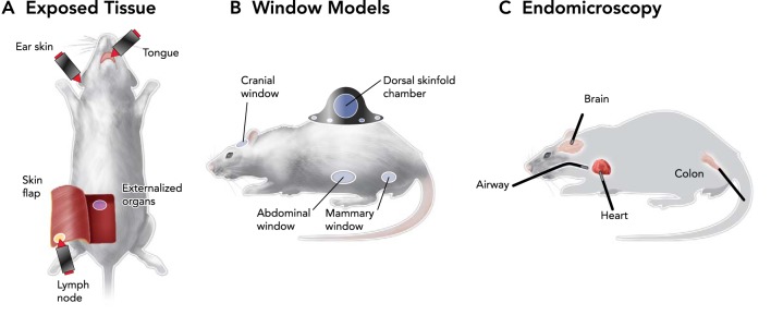
Ways to obtain microscopic access to various organs in vivo Three strategies for intravital imaging have been proposed to overcome limited penetration of light into tissue: through intact or surgically exposed tissue (A), by implanting an optically transparent window (B), or by inserting miniature endoscopic probes (C).
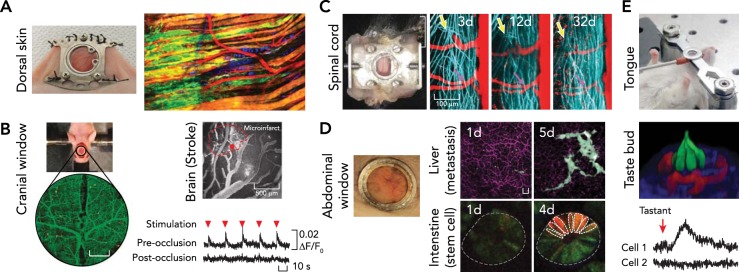
Imaging through optical windows A: dorsal skin. The photograph at left shows a dorsal skinfold chamber implanted on a nude mouse. The fluorescent image at right shows collective invasion of melanoma cells taken by multi-photon microscopy in the dorsal skinfold chamber. Images were reproduced from Ref. with permission. B: brain. The photograph shows cranial window model implanted on a nude mouse after craniotomy. Change in neuronal responsiveness to limb stimulation (red arrowhead) after laser-induced microinfarct. Images reproduced from Ref. with permission. C: spinal cord. The photograph at left shows imaging chamber implanted for chronic imaging of the spinal cord. Regeneration dynamics of the spinal axons (blue) at the injured area (yellow arrow) was traced over a month. Images reproduced from Ref. with permission. D: abdominal organs. Glass window implanted on the abdomen (left) enabled tracking of cellular dynamics in various abdominal organs over time, such as cancer metastasis in the liver (top) and stem cell dynamics in small intestine (bottom). Scale bar, 20 μm. Images reproduced from Refs. – with permission. E: tongue. Top: preparation for imaging the mouse tongue. The fluorescence image (middle) shows a three-dimensional view of a taste bud (red, blood vessel; green, taste receptor cells). Bottom: intracellular calcium activity measured in the taste receptor cells after introducing artificial sweetener intravenously. Images reproduced with permission from unpublished observations of Choi M, Lee WM, Yun SH.
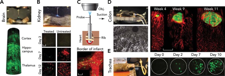
Endomicroscopy A: brain. A side-viewing probe is inserted down to thalamus to visualize neurons labeled by fluorescent protein. Images reproduced from Ref. with permission. B: kidney. Tracking of immune cells over 2 wk was shown with endomicroscopy of the kidney after minimal laparotomy. Scale bar, 50 μm. Image reproduced from Ref. with permission. C: heart. Endomicroscopy with a suction stabilizer was inserted through the intercostal space to access heart with minimal invasiveness for serial imaging over a day. Scale bar, 50 μm. Images reproduced from Ref. with permission. D: colon. Endomicroscopy for the colon. Tumorigenesis and angiogenesis were traced with dual-color endomicroscopic imaging in the descending colon. Scale bar, 500 μm. Images reproduced from Ref. with permission. E: trachea. Endomicroscope inserted through the trachea showed regeneration dynamics of epithelial cells after injury. Scale bar, 20 μm. Images reproduced from Ref. with permission.
References
-
- Akiyoshi T, Lee W, Chase C, Alessandrini A, Sebastian D, Della Pelle P, Connoly S, Farkash E, Yun SH, Russell P. In vivo two photon microscopy of aortic allografts: a new tool for investigation of the dynamics of graft vascular rejection. Am J Transplant 12: 466, 2012.
-
- Alivisatos AP, Andrews AM, Boyden ES, Chun M, Church GM, Deisseroth K, Donoghue JP, Fraser SE, Lippincott-Schwartz J, Looger LL, Masmanidis S, McEuen PL, Nurmikko AV, Park H, Peterka DS, Reid C, Roukes ML, Scherer A, Schnitzer M, Sejnowski TJ, Shepard KL, Tsao D, Turrigiano G, Weiss PS, Xu C, Yuste R, Zhuang X. Nanotools for neuroscience and brain activity mapping. ACS Nano 7: 1850–1866, 2013. - PMC - PubMed
Publication types
MeSH terms
Grants and funding
LinkOut - more resources
Full Text Sources
Other Literature Sources