. 2015 Feb;93(2):165-76.
doi: 10.1007/s00109-014-1247-x. Epub 2015 Jan 7.

Animal model of Sar1b deficiency presents lipid absorption deficits similar to Anderson disease

Affiliations

Animal model of Sar1b deficiency presents lipid absorption deficits similar to Anderson disease

Daniel S Levic et al. J Mol Med (Berl). 2015 Feb.

Abstract

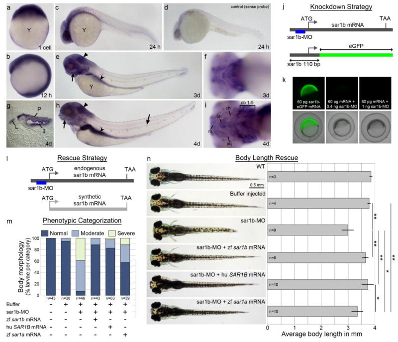
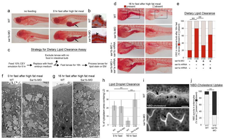
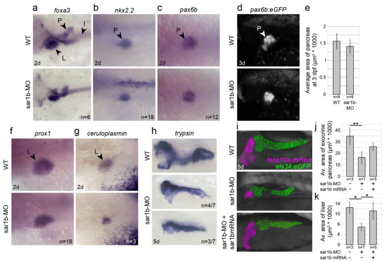
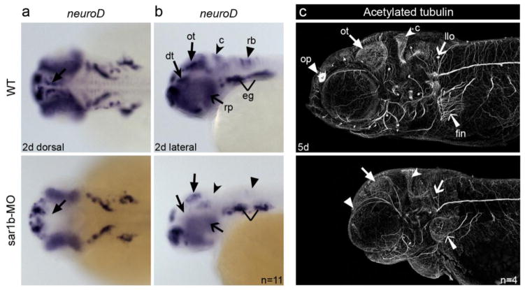
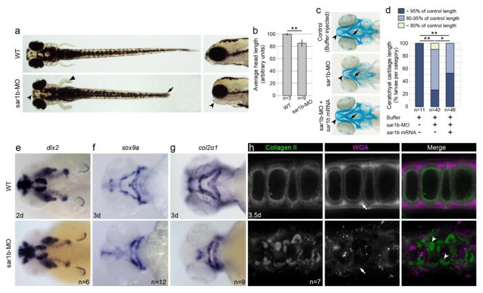
Anderson disease (ANDD) or chylomicron retention disease (CMRD) is a rare, hereditary lipid malabsorption syndrome associated with mutations in the SAR1B gene that is characterized by failure to thrive and hypocholesterolemia. Although the SAR1B structure has been resolved and its role in formation of coat protein II (COPII)-coated carriers is well established, little is known about the requirement for SAR1B during embryogenesis. To address this question, we have developed a zebrafish model of Sar1b deficiency based on antisense oligonucleotide knockdown. We show that zebrafish sar1b is highly conserved among vertebrates; broadly expressed during development; and enriched in the digestive tract organs, brain, and craniofacial skeleton. Consistent with ANDD symptoms of chylomicron retention, we found that dietary lipids in Sar1b-deficient embryos accumulate in enterocytes. Transgenic expression analysis revealed that Sar1b is required for growth of exocrine pancreas and liver. Furthermore, we found abnormal differentiation and maturation of craniofacial cartilage associated with defects in procollagen II secretion and absence of select, neuroD-positive neurons of the midbrain and hindbrain. The model presented here will help to systematically dissect developmental roles of Sar1b and to discover molecular and cellular mechanisms leading to organ-specific ANDD pathology. Key messages: Sar1b depletion phenotype in zebrafish resembles Anderson disease deficits. Sar1b deficiency results in multi-organ developmental deficits. Sar1b is required for dietary cholesterol uptake into enterocytes.


Conflict of interest statement

DISCLOSURE

The authors declare that they do not have any competing or financial interests.

Figures

Figure 1
Figure 1. Sar1b is expressed in select neural tissues, pharyngeal arches and gut
Sar1b mRNA expression by whole mount in situ hybridization at 1cell stage (a), 12 hpf (b), 24 hpf (c), 3 dpf (e, f) and 4 dpf (g–i). Embryos are oriented with head towards the left in lateral views (c, d, e, h), ventral views (f, i) and sagittal section of the mid-gut region at 4 dpf (g). At 24 hpf, expression is concentrated within the ventral brain region (c) and not detected by the sense probe (d). Pigment cells appear as dark spots in C and D. By 3 dpf, transcripts are present throughout the brain, eye, lateral line organ (flat arrow) and enriched in pharyngeal arches (concave arrow) gut (concave arrowhead), and cerebellar plate (flat arrowhead) (e). By 4 dpf the expression persists in the same locations (h), including intestinal epithelium, pancreas, and liver (g), as well as the pharyngeal arches (i). Targeting efficiency of sar1b-MO (J–K). (j) Schematic representation of the knockdown strategy showing sar1b mRNA and position of the translation blocking morpholino (sar1b-MO). The sar1b 5′UTR-eGFP fusion protein mRNA construct contains the sar1b-MO binding site. (k) Double injected embryos with fusion construct and increasing doses of sar1b-MO show almost complete knockdown of GFP expression at 0.4 ng (middle panel) and complete absence of GFP expression at 1 ng of sar1b-MO (right panel). Targeting specificity of sar1b-MO (L–O). (l) Schematic representation of the rescue strategy showing sar1b mRNA and synthetic sar1b mRNA, which lacks the sar1b-MO binding site and which is used for rescue experiments. (m) Double-blinded phenotypic categorization of overall morphology at 4 dpf according to body length, head size, and pectoral fin shape. 54.3% of sar1b-MO injected embryos exhibit a moderate phenotype (representative example in panel N), while 88.4% of embryos co-injected with sar1b-MO and sar1b mRNA are morphologically normal at 4 dpf. Human (hu) SAR1B mRNA rescues morphogenesis similarly to zebrafish (zf) sar1b, while zf sar1a mRNA partially compensates for sar1b knockdown. (n) Images of live embryos with body length quantification for rescue experiments. Sar1b morphants exhibit reduced body length while sar1b-MO + zf sar1b mRNA or hu SAR1B mRNA co-injected embryos do not have significant reduction of body length at 4 dpf. zf sar1a partially rescues body length in sar1b-MO injected embryos. Error bars are ± SD. Abbreviations: y, yolk, m, Meckel’s cartilage, bh, basihyal, pq, palatoquadrate, ch, ceratohyal, cb, ceratobranchials 1–5, I, intestine, L, liver, P, pancreas; zf, zebrafish; hu, human. * p < 0.05, ** p < 0.01; One-way ANOVA standard weighted means analysis with Tukey’s test.
Figure 2
Figure 2. Sar1b knockdown impairs dietary lipid absorption and cholesterol uptake
(a) Whole-mount detergent-free Oil Red O (ORO) staining of WT and sar1b morphant embryos under non-fed fasting conditions (left) and immediately after feeding a high fat meal (10% chicken egg yolk) [36]. After feeding, the intestinal bulb contains abundant lipids (arrow). (b) ORO staining of gut cryosections after feeding shows that lipids accumulate within enterocytes (concave arrowhead). (c) Schematic of experimental design and workflow for testing dietary lipid clearance in zebrafish larvae. Sar1b knockdown impairs dietary lipid clearance from enterocytes (D–H). (d) ORO staining of WT, sar1b-MO, and sar1b-MO + mRNA co-injected larvae fasted 16 hours after feeding. Left panels show representative examples of larvae with lipids remaining in the gut after fasting (present), while right panels show larvae that have cleared lipids from the intestinal bulb (absent). (e) Quantification of ORO presence in intestinal bulb after lipid clearance assay. Sar1b knockdown significantly impairs dietary lipid clearance, which can be rescued with sar1b mRNA expression. Sar1a mRNA does not effectively compensate for sar1b knockdown. (f) Transmission electron microscopy (TEM) analysis of WT and sar1b morphant larvae immediately after feeding 10% CEY. Lipid droplets (LD) are present in the enterocyte cytoplasm after a high fat meal [37]. Scale bars represent 5 μm. (g) TEM of WT and sar1b morphant larvae after lipid clearance assay. WT enterocytes contain few LDs compared to sar1b morphants [38, 39]. Scale bars represent 5 μm. (h) Quantification of lipid droplet abundance in enterocyte cytoplasm from TEMs. (i) NBD-cholesterol is present in the intestinal lumen of WT and morphants (arrows) when fed a high fat meal spiked with the cholesterol-fluorophore conjugate, but cholesterol positive inclusions are present only in the WT enterocytes (arrowheads) and absent in 71.4% of Sar1b morphants. Scale bars represent 20 μm. (j) Quantification of NBD fluorescence in enterocytes after feeding. ** p<0.01, Fisher’s exact test (E, J) or two way ANOVA with Tukey’s test (H). Error bars are ± SD. Abbreviations: BB, brush border; LD, lipid droplet; N, nucleus.
Figure 3
Figure 3. Sar1b knockdown results in reduced size of digestive organs
(a–c) Transcripts of foxa3, nkx2.2, and pax6b detected by whole-mount in situ hybridization show no significant differences in spatiotemporal patterning and specification of pancreatic precursors at 2 dpf. (d–e) Expression of the (pax6b:GFP) transgene shows modest but insignificant reduction in pancreas size at 3 dpf. (f–g) Transcripts of prox1 and ceruloplasmin show no significant differences in spatiotemporal patterning and specification of liver precursors at 2 dpf. (h) Transcripts of trypsin show reductions in pancreas size and change in lobular shape at 5 dpf. (i–k) Expression of the liver (fabp10a:dsRed) and pancreas (ela3A:eGFP) transgenes show consistently reduced size in 5 dpf Sar1b morphants, which can be partially rescued with -sar1b mRNA expression. Error bars are ± SD. Abbreviations: L, liver, P, pancreas, I, intestinal bulb. * p < 0.05, ** p < 0.01; two-tailed unpaired t-test (E) or One-way ANOVA standard weighted means analysis with Tukey’s test (J–K).
Figure 4
Figure 4. Depletion of Sar1b disrupts neural development
(a, b) Whole mount in situ hybridization with riboprobe detecting neuroD transcripts in 2 dpf embryos shows relatively normal spatio-temporal expression in epibranchial ganglia (eg), but reduced or absent expression demarcating neuroprogenitors in the dorsal thalamus (dt), optic tectum (ot), cerebellum (c) and hindbrain rhombomeres (rb), as well as retinal progenitors (rp). Dorsal (a) and (b) lateral views. (c) Maximum intensity z-projections of acetylated tubulin stained axonal tracks reveals reduction in staining in the olfactory pits (op), lateral line (llo) sensory patches of the head and pectoral fin innervation (fin). The forebrain and the optic tectum (ot) are reduced in size and shifted anteriorly in the morphants.
Figure 5
Figure 5. Sar1b knockdown results in skeletal dysmorphology
(a) Live images of WT and sar1b morphants show shorter head in anteroposterior direction (concave arrowhead), kinked pectoral fins (flat arrowhead) and a shorter overall body length (arrow) in dorsal and lateral head views. (b) Quantification of anteroposterior head length measurements at 4 dpf. (c) Alcian blue stained head skeleton preparations revealed a shortened Meckel’s (concave arrowhead) and malformed ceratohyal (Ch) cartilage elements (arrow) of Sar1b-MO compared to control embryos, while sar1b-MO + sar1b mRNA co-injection rescued cartilage morphology. (d) Quantification of Ch cartilage length of Alcian blue stained embryos. Control length refers to the average length of buffer-injected embryos. (e) Unchanged dlx2 expression in Sar1b-MO shows that neural crest cell migration and patterning are normal in sar1b morphant embryos at 2 dpf. (f) Chondrogenic differentiation marker sox9a, (g) and its target gene col2a1 show reduced size and malformed morphant cartilages compared to WT. (h) Immunofluorescence staining with antibody recognizing Collagen II and of N-glycans using wheat germ agglutinin (WGA) shows intracellular inclusions of procollagen type II (arrowheads) and reduced staining intensity of N-glycans within extracellular matrix (arrows). Error bars are ± SD. * p < 0.05, ** p < 0.01; two-tailed unpaired t-test (b) or Fisher’s exact test (d).

Comment in

Similar articles

Cited by

References

    1. Abumrad NA, Davidson NO. Role of the Gut in Lipid Homeostasis. Physiol Rev. 2012;92:1061–1085. - PMC - PubMed
    1. Iqbal J, Hussain MM. Intestinal lipid absorption. Am J Physiol - Endocrinol Metab. 2009;296:E1183–E1194. - PMC - PubMed
    1. Miller EA, Barlowe C. Regulation of coat assembly—sorting things out at the ER. Curr Opin Cell Biol. 2010;22:447–453. - PMC - PubMed
    1. Siddiqi SA, Gorelick FS, Mahan JT, Mansbach CM. COPII proteins are required for Golgi fusion but not for endoplasmic reticulum budding of the pre-chylomicron transport vesicle. J Cell Sci. 2003;116:415–427. - PubMed
    1. Walther TC, Farese RV. Lipid Droplets and Cellular Lipid Metabolism. Annu Rev Biochem. 2012;81:687–714. - PMC - PubMed

Publication types

MeSH terms

Substances

Supplementary concepts