. 2015:4:158-68.
doi: 10.1016/j.redox.2014.12.009. Epub 2014 Dec 24.

Steatosis-induced proteins adducts with lipid peroxidation products and nuclear electrophilic stress in hepatocytes

Affiliations

Steatosis-induced proteins adducts with lipid peroxidation products and nuclear electrophilic stress in hepatocytes

Sarit Anavi et al. Redox Biol. 2015.

Abstract

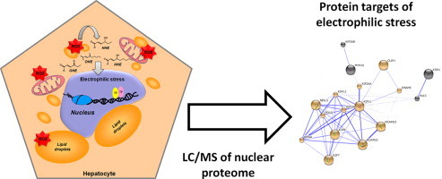
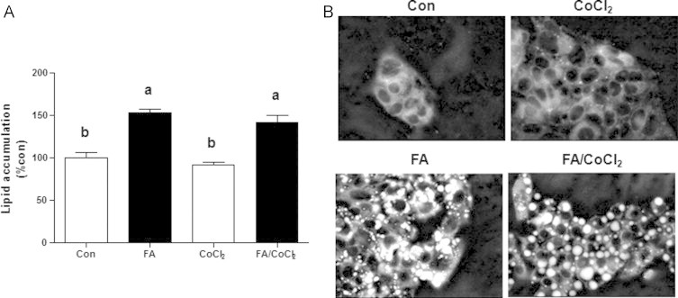
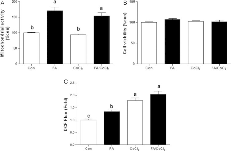
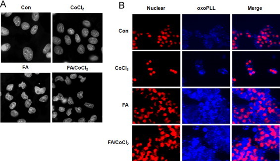
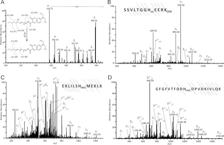
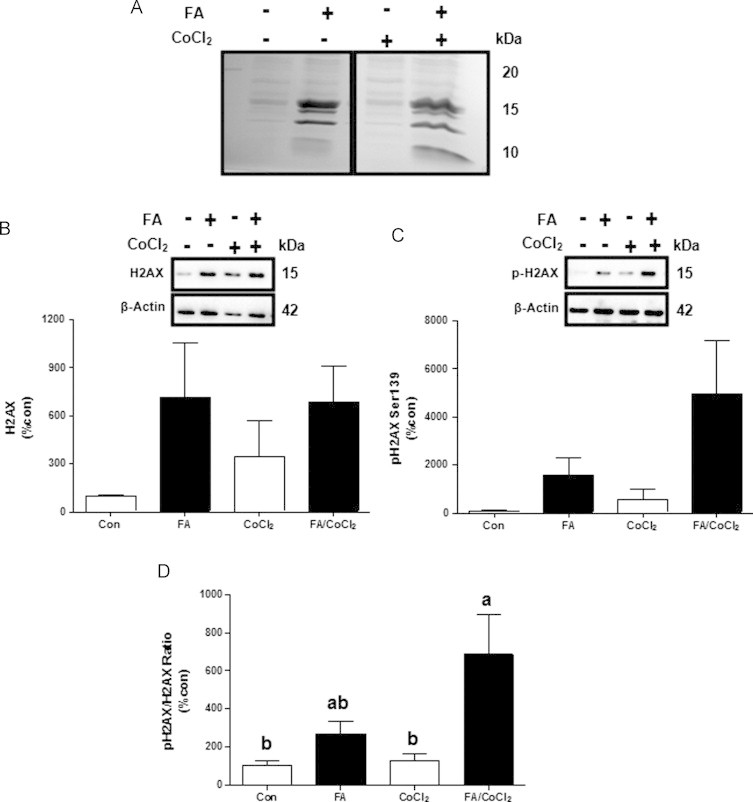
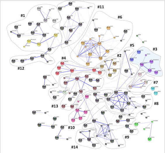
Accumulating evidence suggests that fatty livers are particularly more susceptible to several pathological conditions, including hepatic inflammation, cirrhosis and liver cancer. However the exact mechanism of such susceptibility is still largely obscure. The current study aimed to elucidate the effect of hepatocytes lipid accumulation on the nuclear electrophilic stress. Accumulation of intracellular lipids was significantly increased in HepG2 cells incubated with fatty acid (FA) complex (1mM, 2:1 oleic and palmitic acids). In FA-treated cells, lipid droplets were localized around the nucleus and seemed to induce mechanical force, leading to the disruption of the nucleus morphology. Level of reactive oxygen species (ROS) was significantly increased in FA-loaded cells and was further augmented by treatment with moderate stressor (CoCl2). Increased ROS resulted in formation of reactive carbonyls (aldehydes and ketones, derived from lipid peroxidation) with a strong perinuclear accumulation. Mass-spectroscopy analysis indicated that lipid accumulation per-se can results in modification of nuclear protein by reactive lipid peroxidation products (oxoLPP). 235 Modified proteins involved in transcription regulation, splicing, protein synthesis and degradation, DNA repair and lipid metabolism were identified uniquely in FA-treated cells. These findings suggest that steatosis can affect nuclear redox state, and induce modifications of nuclear proteins by reactive oxoLPP accumulated in the perinuclear space upon FA-treatment.

Keywords: Electrophilic stress; Fatty liver diseases; Lipid peroxidation; Protein modifications; Reactive carbonyls.


Figures

None
Graphical abstract
Fig. 1
Fig. 1
Effects of fatty acids treatment and/or CoCl2 on cellular lipids accumulation in HepG2 cells. Cells were incubated with 1 mM fatty acid mixture (oleate/palmitate, 2/1; black bars) or vehicle in which fatty acids were dissolved (white bars) for 24 h. Afterwards, cells were treated with or without 100 µM CoCl2 for 4 h. (A) Intracellular lipid levels were evaluated quantitatively and qualitatively using Nile red staining and flow cytometry analysis or (B) fluorescence microscopy. Results are means±SE. Means with different letters in each group differ at p<0.05. Con, control; FA, fatty acids.
Fig. 2
Fig. 2
Effects of fatty acids treatment and/or CoCl2 on mitochondrial activity and ROS production. Cells were incubated with 1 mM fatty acid mixture (oleate/palmitate, 2/1; black bars) or vehicle in which fatty acids were dissolved (white bars) for 24 h. Afterwards, cells were treated with or without 100 µM CoCl2 for 4 h. (A) Mitochondrial activity was evaluated using MTT assay and (B) cell viability was assessed using neutral-red staining. (D) ROS levels following 3.5 h of incubation with or without CoCl2 in normal and FA-loaded cells measured by DCFDA assay. Results are means±SE. Means with different letters in each group differ at p<0.05. Con, control; FA, fatty acids.
Fig. 3
Fig. 3
Effects of fatty acids treatment and/or CoCl2 on nuclear morphology and lipid peroxidation products (oxoLPP) formation. Cells were incubated with 1 mM fatty acid mixture (oleate/palmitate; 2/1) or vehicle in which fatty acids were dissolved for 24 h. Afterwards, cells were treated with or without 100 µM CoCl2 for 4 h. (A) Nuclear morphology and (B) oxoLPP spatial distribution were observed using fluorescence microscopy. Con, control; FA, fatty acids.
Fig. 4
Fig. 4
LTQ CID spectra of carbonylated LPP and oxoLPP modified peptides. For detection of oxoLPP cell pellets were collected, derivatized with 7-(diethylamino)coumarin-3-carbohydrazide (CHH), lipids were extracted and analyzed by direct infusion ESI-LTQ-Orbitrap. (A) LTQ CID spectrum of CHH-derivatized 4-hydroxy-nonenal. For identification of oxoLPP-modified proteins, nuclear proteins were separated by SDS-PAGE, in-gel digested with trypsin and analyzed by nUPLC-ESI-LTQ-Orbitrap MS. LTQ CID spectra of peroxisome-proliferator-activated receptor-γ coactivator 1α OHE-modified peptide 361SSVLTGGHoxEERKOHE371 (B), histone acetyltransferases KAT6B HHE-modified peptide 961EKLILSHHHEMEKLK972 (C), and hnRNP A/B HHE-modified peptide 154GFGFVTFDDHHNEDPVDKIVLQK173 (D).
Fig. 5
Fig. 5
Effects of fatty acids treatment and/or CoCl2 on histone H2AX. Cells were incubated with 1 mM fatty acid mixture (oleate/palmitate, 2/1; black bars) or vehicle in which fatty acids were dissolved (white bars) for 24 h. Afterwards, cells were treated with or without 100 µM CoCl2 for 4 h. (A) Representative SDS PAGE gels and (B) total and (C) phosphorylation status of histone 2AX (H2AX) at Ser139. (D) H2AX total to phosphorylated ratio. Results are means±SE. Means with different letters in each group differ at p<0.05. Con, control; FA, fatty acids.
Fig. 6
Fig. 6
STRIG protein–protein interaction and clustering analysis of oxoLPP modified proteins identified in fatty acids treated HepG2 cells. 332 oxoLPP modified proteins identified in FA group by LC–MS analysis were uploaded to STRIG database of protein–protein interaction. Resulted network was clustered based on the Markov cluster algorithm (MCL), disconnected nodes were removed and resulted clusters were manually annotated. Corresponding cluster numbering legend is provided in Table 1.

References

    1. Feldstein A.E., Bailey S.M. Emerging role of redox dysregulation in alcoholic and nonalcoholic fatty liver disease. Antioxidants & Redox Signaling. 2011;15(2):421–424. 21254858 - PMC - PubMed
    1. Hansen J.M., Go Y.M., Jones D.P. Nuclear and mitochondrial compartmentation of oxidative stress and redox signaling. Annual Review of Pharmacology and Toxicology. 2006;46:215–234. 16402904 - PubMed
    1. Lukosz M., Jakob S., Büchner N., Zschauer T.C., Altschmied J., Haendeler J. Nuclear redox signaling. Antioxidants & Redox Signaling. 2010;12(6):713–742. 19737086 - PubMed
    1. Strand J.M., Scheffler K., Bjørås M., Eide L. The distribution of DNA damage is defined by region-specific susceptibility to DNA damage formation rather than repair differences. DNA Repair. 2014;18:44–51. 24685126 - PubMed
    1. Shlomai J. Redox control of protein–DNA interactions: from molecular mechanisms to significance in signal transduction, gene expression, and DNA replication. Antioxidants & Redox Signaling. 2010;13(9):1429–1476. 20446770 - PubMed

Publication types