Review
. 2015 Mar;11(3):171-81.
doi: 10.1038/nrendo.2014.226. Epub 2015 Jan 6.

TSH-receptor-expressing fibrocytes and thyroid-associated ophthalmopathy

Affiliations
Review

TSH-receptor-expressing fibrocytes and thyroid-associated ophthalmopathy

Terry J Smith. Nat Rev Endocrinol. 2015 Mar.

Abstract

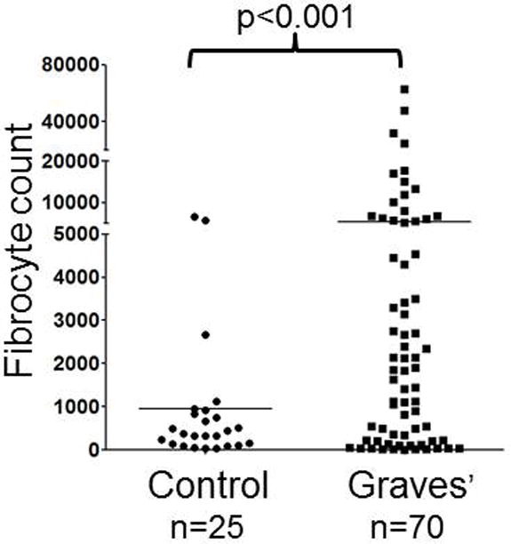
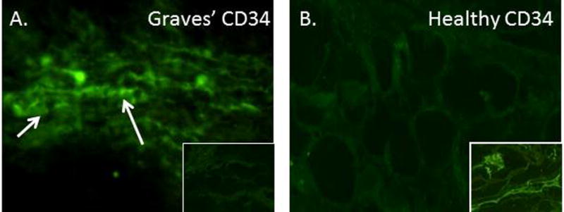
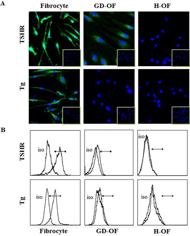
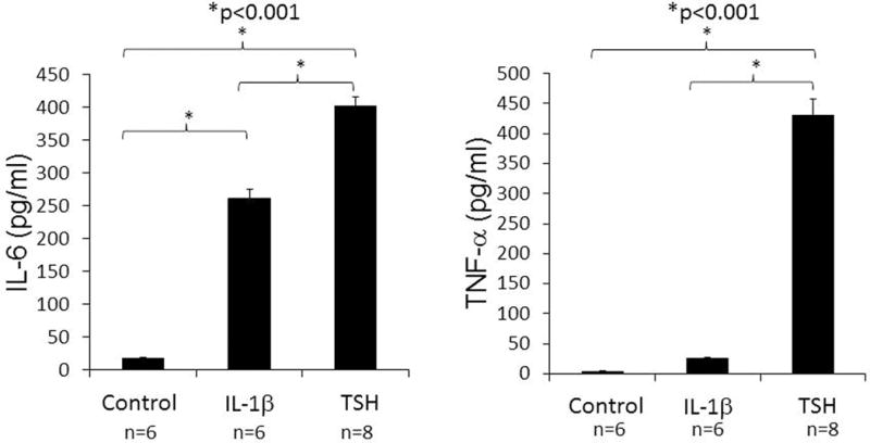
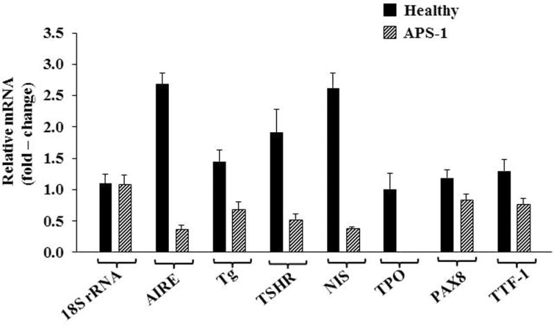
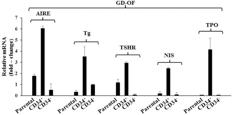
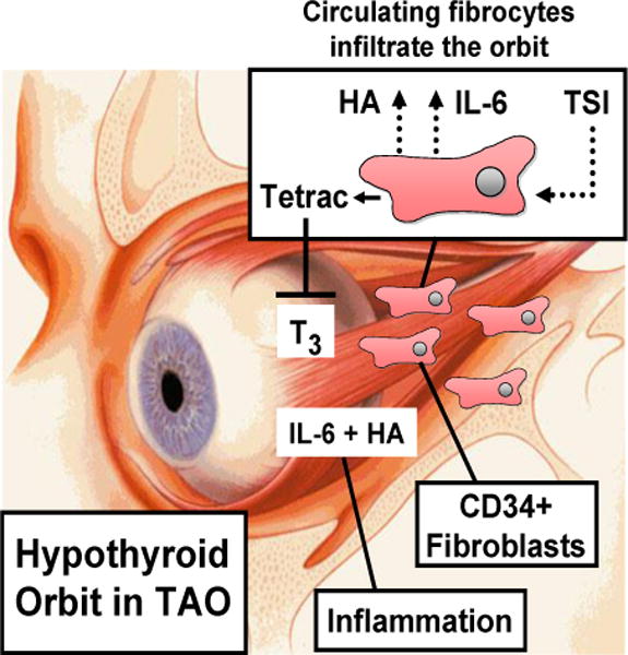
Thyroid-associated ophthalmopathy (TAO) is a vexing and undertreated ocular component of Graves disease in which orbital tissues undergo extensive remodelling. My colleagues and I have introduced the concept that fibrocytes expressing the haematopoietic cell antigen CD34 (CD34(+) fibrocytes), which are precursor cells of bone-marrow-derived monocyte lineage, express the TSH receptor (TSHR). These cells also produce several other proteins whose expression was traditionally thought to be restricted to the thyroid gland. TSHR-expressing fibrocytes in which the receptor is activated by its ligand generate extremely high levels of several inflammatory cytokines. Acting in concert with TSHR, the insulin-like growth factor 1 receptor (IGF-1R) expressed by orbital fibroblasts and fibrocytes seems to be necessary for TSHR-dependent cytokine production, as anti-IGF-1R blocking antibodies attenuate these proinflammatory actions of TSH. Furthermore, circulating fibrocytes are highly abundant in patients with TAO and seem to infiltrate orbital connective tissues, where they might transition to CD34(+) fibroblasts. My research group has postulated that the infiltration of fibrocytes into the orbit, their unique biosynthetic repertoire and their proinflammatory and profibrotic phenotype account for the characteristic properties exhibited by orbital connective tissues that underlie susceptibility to TAO. These insights, which have emerged in the past few years, might be of use in therapeutically targeting pathogenic orbit-infiltrating fibrocytes selectively by utilizing novel biologic agents that interfere with TSHR and IGF-1R signalling.


Conflict of interest statement

The author affirms no competing interests.

Figures

Figure 1
Figure 1
Increased frequency of CD34+ fibrocyte generation from the PBMCs of 70 patients with GD compared with 25 healthy control donors. PBMCs were inoculated at a density of 5 × 10⁶ cells/well. Cultures were incubated for 14d. Adherent cells (<5% of starting cells) were collected and counted. (Reprinted with permission; Douglas et al. Increased generation of fibrocytes in thyroid-associated ophthalmopathy, Reference 6, Copyright 2010. The Endocrine Society.)
Figure 1
Figure 1
Increased frequency of CD34+ fibrocyte generation from the PBMCs of 70 patients with GD compared with 25 healthy control donors. PBMCs were inoculated at a density of 5 × 10⁶ cells/well. Cultures were incubated for 14d. Adherent cells (<5% of starting cells) were collected and counted. (Reprinted with permission; Douglas et al. Increased generation of fibrocytes in thyroid-associated ophthalmopathy, Reference 6, Copyright 2010. The Endocrine Society.)
Figure 2
Figure 2
CD34+ LSP-1+ TSHR+ fibrocytes can be identified in the orbital tissue of patients with TAO but are absent in tissues from healthy donors. A, CD34 expression (arrows, green fluorescein isothiocyanate) in TAO- derived tissue (inset, negative control staining). B, Absent, CD34 expression in healthy tissue (inset, positive control). C, LSP-1 expression in TAO-derived tissue [red, arrows, nuclei counter-stained with 4′,6′ –diamino-2-phenylindole (DAPI)(blue)(inset, negative, control). D, Absence of LSP-1 expression in healthy tissue (inset, negative controls). E, CD31 expression in disease-derived tissue is limited to vascular endothelium (red, arrows). F, Hematoxylin and eosin-stained consecutive thin sections of the same orbital tissue (×40). G, Fibrocytes present in orbital tissue from patients with TAO co-express CD34 and TSHR. Thin-sectioned tissue from a donor with TAO was stained with anti-CD34 (green) and anti-TSHR (red) antibodies. Nuclei were counterstained with DAPI (blue). Thin sections were then subjected to confocal microscopy. Inset contains a negative staining control. (Reprinted with permission; Douglas et al. Increased generation of fibrocytes in thyroid-associated ophthalmopathy, Reference 6, Copyright 2010. The Endocrine Society.)
Figure 3
Figure 3
Analysis of TSHR and Tg expression by human fibrocytes and fibroblasts by (A) immunofluorescence staining and (B) flow cytometry. Cultivated cells were (A) fixed, blocked, and stained with either anti-TSHR, anti-Tg, or isotype control mouse IgG followed by Alexa Fluor 488-conjugated goat anti-mouse Abs. Nuclei were stained with DAPI, and thin sections were subjected to confocal fluorescence microscopy. (Insets) Isotype IgG staining. (B) Fibrocytes and fibroblasts were incubated with phycoerythrin-conjugated anti-TSHR, anti-TG, or isotype mAbs and Alexa Fluor 488 goat anti-mouse IgG. They were subjected to flow cytometry. Horizontal lines denote fluorescence intensity compared with isotype controls. (Reprinted from; Fernando et al. Human fibrocytes co-express thyroglobulin and thyrotropin receptor, Proceedings of the National Academy of Sciences, Reference 103, Copyright 2012.)
Figure 4
Figure 4
TSHR displayed on fibrocytes generated from PBMCs can initiate cytokine production. Cultures cells from a patient with GD were treated with bTSH (5 mU/ml) or IL-1β (10 ng/ml) for 48h. Medium was subjected to ELISAs specific for IL-6 (left panel) or TNF- α (right panel). Data are expressed as the mean ± SEM of three replicate culture wells. *, P < 0.001. (Reprinted with permission; Douglas et al. Increased generation of fibrocytes in thyroid-associated ophthalmopathy, Reference 6, Copyright 2010. The Endocrine Society.)
Figure 4
Figure 4
TSHR displayed on fibrocytes generated from PBMCs can initiate cytokine production. Cultures cells from a patient with GD were treated with bTSH (5 mU/ml) or IL-1β (10 ng/ml) for 48h. Medium was subjected to ELISAs specific for IL-6 (left panel) or TNF- α (right panel). Data are expressed as the mean ± SEM of three replicate culture wells. *, P < 0.001. (Reprinted with permission; Douglas et al. Increased generation of fibrocytes in thyroid-associated ophthalmopathy, Reference 6, Copyright 2010. The Endocrine Society.)
Figure 5
Figure 5
Interfering with AIRE expression knocked-down levels of AIRE, Tg, TSHR, NIS, TPO, PAX8, and TTF-1 but not PGHS-2 or 18S RNA. A, Fibrocytes from four different donors were transfected, either with scrambled (control) siRNA or siRNA directed against AIRE. Cultures were then incubated for 48 hours, and RNA was harvested and then subjected to real-time PCR. Data are expressed as mean ± SD of three independent determinants. B, Fibrocyte cultures were transfected with either siRNA or siRNA targeting AIRE for 96 hours. Tg protein was quantified by labeling with [35S]methionine (40μCi/mL) and then immunoprecipitated. TSHR and NIS were quantified by flow cytometry. Levels of TPO were assessed by Western blotting. (Reprinted with permission; Fernando et al. Expression of thyrotropin receptor, thyroglobulin, sodium iodide symporter, and thyroperoxidase by fibrocytes depends on AIRE, Reference 104, Copyright 2014. The Endocrine Society.)

References

    1. Brent GA. Graves’ Disease. N Engl J Med. 2008;358:2594–2605. - PubMed
    1. Bahn RS. Mechanisms of Disease Graves’ Ophthalmopathy. N Engl J Med. 2010;362:726–738. - PMC - PubMed
    1. Rundle FF, Wilson CW. Development and course of exophthalmos and ophthalmoplegia in Graves’ disease with special reference to the effect of thyroidectomy. Clin Sci. 1945;5:177–194. - PubMed
    1. Smith TJ, Bahn RS, Gorman CA. Connective tissue, glycosaminoglycans, and diseases of the thyroid. Endocr Rev. 1989;10:366–391. - PubMed
    1. Smith TJ, Tsai CC, Shih JJ, Tsui S, et al. Unique attributes of orbital fibroblasts and global alterations in IGF-1 receptor signaling could explain thyroid-associated ophthalmopathy. Thryoid. 2008;18:983–988. - PMC - PubMed

Publication types