. 2015 Mar;167(3):854-71.
doi: 10.1104/pp.114.255430. Epub 2015 Jan 5.

Network of GRAS transcription factors involved in the control of arbuscule development in Lotus japonicus

Affiliations

Network of GRAS transcription factors involved in the control of arbuscule development in Lotus japonicus

Li Xue et al. Plant Physiol. 2015 Mar.

Abstract

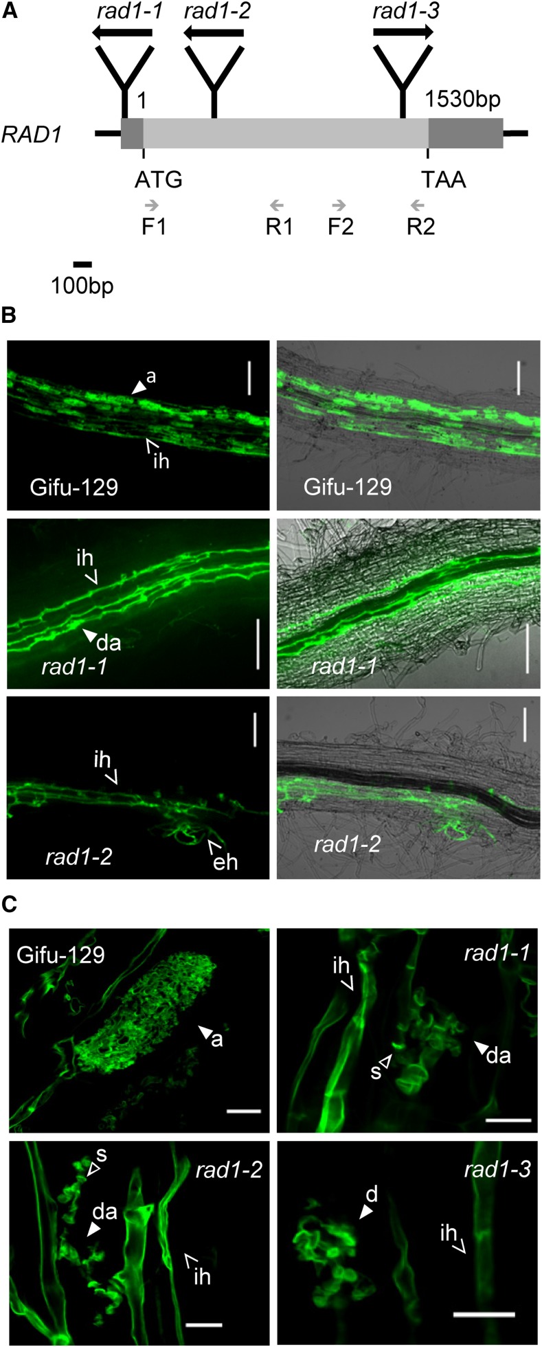
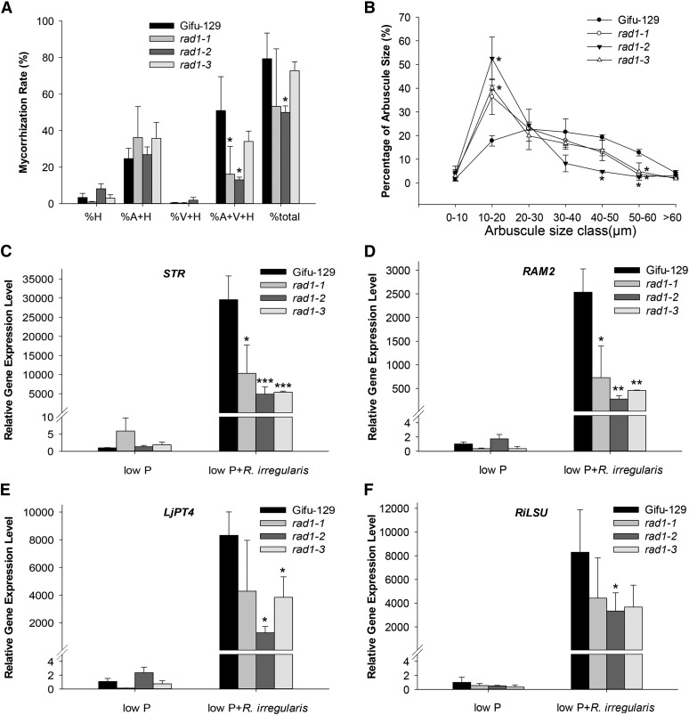
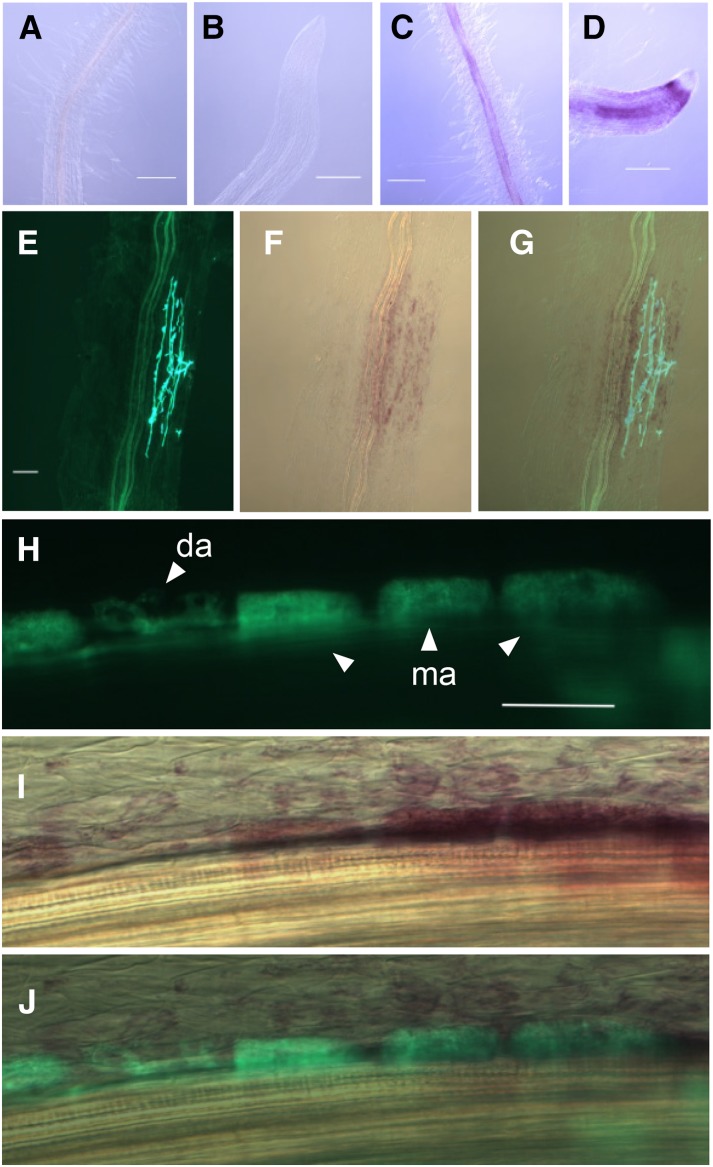
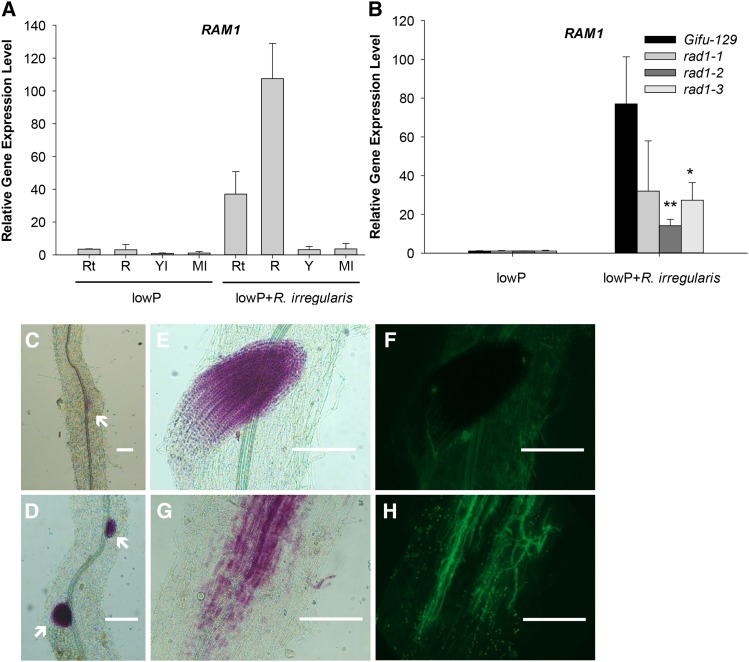
Arbuscular mycorrhizal (AM) fungi, in symbiosis with plants, facilitate acquisition of nutrients from the soil to their host. After penetration, intracellular hyphae form fine-branched structures in cortical cells termed arbuscules, representing the major site where bidirectional nutrient exchange takes place between the host plant and fungus. Transcriptional mechanisms underlying this cellular reprogramming are still poorly understood. GRAS proteins are an important family of transcriptional regulators in plants, named after the first three members: GIBBERELLIC ACID-INSENSITIVE, REPRESSOR of GAI, and SCARECROW. Here, we show that among 45 transcription factors up-regulated in mycorrhizal roots of the legume Lotus japonicus, expression of a unique GRAS protein particularly increases in arbuscule-containing cells under low phosphate conditions and displays a phylogenetic pattern characteristic of symbiotic genes. Allelic rad1 mutants display a strongly reduced number of arbuscules, which undergo accelerated degeneration. In further studies, two RAD1-interacting proteins were identified. One of them is the closest homolog of Medicago truncatula, REDUCED ARBUSCULAR MYCORRHIZATION1 (RAM1), which was reported to regulate a glycerol-3-phosphate acyl transferase that promotes cutin biosynthesis to enhance hyphopodia formation. As in M. truncatula, the L. japonicus ram1 mutant lines show compromised AM colonization and stunted arbuscules. Our findings provide, to our knowledge, new insight into the transcriptional program underlying the host's response to AM colonization and propose a function of GRAS transcription factors including RAD1 and RAM1 during arbuscule development.


Figures

Figure 1.
Figure 1.
Global analysis of mycorrhiza up-regulated transcription factors. A, Hierarchical clustering of mycorrhizal (+M) and nonmycorrhizal wild-type Gifu-129 expression profiles based on transcript abundance of the genome-wide 45 AM up-regulated transcription factors identified by RNAseq (adjusted P < 0.01, fold change > 2). Gifu-129 plants were grown for 8 weeks at low Pi (5.5 μm; low P) or high Pi (7.5 mm; high P) conditions, respectively, or they were grown at low Pi for 6 weeks with a subsequent shift from low Pi to high Pi for 2 weeks (shift). Significantly up-regulated transcription factors are shown in a heat map drawn by using R. Log10 values of normalized counts per transcript plus one were used. Transcription factor families are labeled above the genes. The closest homologs and RAD1 were labeled below the plot. Asterisks indicate the genes whose expression was confirmed by qRT-PCR in Supplemental Figure S1. B, The 43 AM up-regulated transcription factors were clustered into three groups in AM host and nonhost species. Amino acid sequences of these L. japonicus AM up-regulated transcription factors were utilized for reciprocal blast in AM nonhost Brassicaceae (Capsella rubella, Thellungiella halophila, Brassica rapa, Arabidopsis lyrata, and Arabidopsis [Arabidopsis thaliana]), Lupinus angustifolius, and AM hosts (M. truncatula, Populus trichocarpa, Carica papaya, Gossypium raimondii, and Vitis vinifera). Symbols indicate the presence (+) or absence (−) of the capacity for symbiosis. Detected homologs are labeled with dark blue. Lack of homolog detection was labeled with white.
Figure 2.
Figure 2.
Mycorrhizal phenotype of rad1 allelic mutants. A, RAD1 (chr4.CM1864.540.r2.m) gene structure is shown at scale, and LORE1a insertion sites are illustrated in mutants rad1-1 (−71 bp upstream of the translation start codon [ATG]), rad1-2 (359 bp downstream of start ATG), and rad1-3 (1,379 bp downstream of start ATG). TAA indicates the translation stop codon. Black arrows indicate the 5′ to 3′ direction of the LORE1a transposon sequence. Gray arrows indicate the specific primers used for RT-PCR. Light-gray bar represents the coding region, and dark-gray bars represent the 5′ UTR and 3′ UTR. B, Confocal microscopy images of R. irregularis in the roots of Gifu-129 and the rad1 allelic homozygous mutants. Images at right show the overlay of fluorescence and bright-field images. Scale bars = 100 µm. C, Defective arbuscules in rad1 allelic homozygous mutants. Fungal structures were stained with WGA-Alexa Fluor 488. White arrowheads indicate characteristic mycorrhizal structures: a, arbuscule; ih, intraradical hyphae; eh, extraradical hyphae; da, degenerated arbuscule; and s, septa. Scale bars = 10 µm.
Figure 3.
Figure 3.
Mycorrhizal phenotype and marker gene expression in allelic rad1 mutants. A, Mycorrhization rate of Gifu-129 and three allelic rad1 mutant lines in the presence of R. irregularis at 6 wpi. Four plants per pot served as one biological replicate. Three biological replicates were used. Asterisks indicate significant differences between the wild type and rad1 mutant using Student’s t test (*P < 0.05). Error bars represent sd (n = 3). At least four independent experiments were performed with similar results. Percent total colonization and percentage of different fungal structures in colonized roots as indicated on x axis were counted. H, Hyphae; V, vesicle; A, arbuscule. B, Frequency of arbuscule size classes in the wild-type Gifu-129 and three allelic rad1 mutant lines. Mean value of frequency was from three biological replicates with approximately 90 root segments per genotype. Approximately 400 arbuscules were measured per genotype. Error bars represent sd (n = 3). Student’s t test was utilized between Gifu-129 and rad1 mutant at each size cluster. Asterisks indicate significant differences between frequency values of Gifu-129 and genotypes corresponding to the respective symbol closest to the asterisk (P < 0.05). C to F, Marker gene expression of STR, RAM2, LjPT4, and R. irregularis large subunit (RiLSU) in the presence or absence of R. irregularis under low-Pi conditions. Four plants per pot served as a biological replicate. Mean values of three biological replicates are presented. Error bars represent sd. Asterisks indicate significant differences (Student’s t test, *P ≤ 0.05, **P < 0.01, and ***P < 0.001).
Figure 4.
Figure 4.
Histochemical analysis of proRAD1:GUS expression in mycorrhizal and nonmycorrhizal hairy roots of L. japonicus. A and B, Absence of GUS activity in nonmycorrhizal roots and root tips under high-Pi conditions. Scale bars = 100 µm. C and D, GUS activity in nonmycorrhizal roots and root tips under low-Pi conditions. Scale bars = 100 µm. E to G, GUS activity was observed in arbuscule-containing and adjacent cells. E, Fluorescence image of WGA-Alexa Fluor 488-stained intraradical fungal structures. F, Bright-field image of magenta GUS-stained root sector shown in E. G, Alexa Fluor 488 and bright-field image were merged to show colocalization of intraradical AM fungal hyphae with GUS activity. Scale bar = 10 µm. Fluorescence of Alexa Fluor 488 (H), bright-field image of magenta GUS-stained arbusculated inner cortical cells (I), and merged images (J). Scale bar = 50 µm. da, Developing arbuscule; ma, mature arbuscule.
Figure 5.
Figure 5.
Phylogenetic analysis of GRAS proteins. An unrooted phylogenetic tree was generated based on amino acid alignment including 18 mycorrhiza-inducible GRAS proteins identified in this work (blue dots); 33 GRAS family members from Arabidopsis, symbiotic GRAS MtRAM1, MtDELLA1/MtDELLA2, OsDIP1, MtNSP1, MtNSP2, LjNSP1, LjNSP2 (pink dots); and the RAD1 homologs from dicots (green dots), monocots (light-green dots), lycophytes (orange dot), and liverworts (cyan dots). The alignment of protein sequences was performed using ClustalX, and the phylogenetic tree was constructed by MEGA5 using the Neighbor-Joining algorithm. Bootstrap values were labeled at each node. The RAD1 orthologs (clade I), paralogs (clade II), and RAD1 orthologs in monocots, lycophytes, and liverworts (clade III) with color background were selected based on e values <10−90 in BLAST using reciprocal analysis. Red star indicates the putative genome duplication event.
Figure 6.
Figure 6.
RAD1 interacts with RAM1 in vitro and in vivo. A, Interaction between truncated versions of RAD1 and full-length RAM1. Conserved domains in GRAS proteins are color coded. BD, GAL4 DNA-binding domain; SD, synthetic minimal media based upon yeast (Saccharomyces cerevisiae) nitrogen base supplemented with Glc and amino acids (aa) except those indicated (L, W, H); 3AT, 3-amino-1,2,4-triazole. cGRAS encoding GRAS protein fused to the GAL4 activation domain (AD) was used as a negative control. B, In vitro pull-down assay. GST fusion proteins RAM1 (GST-RAM1) and NSP2 (GST-NSP2) or GST alone (control) were used to precipitate His-tag fused RAD1 (His-RAD1). + indicates the presence of the recombinant protein. WB: anti-His, Western blotting with anti-His antibody detecting His-bait fusion proteins. Purified GST fusion protein through glutathione-Sepharose beads was analyzed by SDS-PAGE and Coomassie Brilliant Blue in-gel protein staining (CBB staining). Arrows at left demonstrate presence of GST fusion proteins as designated. C, In vivo Co-IP of HA-RAD1 by GFP-RAM1. In vivo Co-IP was performed using N. benthamiana leaves infiltrated with A. rhizogenes GV3101 strain harboring the desired interacting pair (HA:RAD1/GFP:RAM1). Leaf extracts were subjected to immunoprecipitation (IP) using anti-GFP antibody. Anti-HA and anti-GFP antibodies were used for western blotting of Co-IP sample. Symbols represent the presence (+) or absence (–) of the various proteins. D, Interaction between RAM1 and RAD1 by BiFC assay in N. benthamiana. Leaves were infiltrated with mixtures of A. tumefaciens strain GV3101 to coexpress RAD1 and RAM1 fused to one-half of split YFP, respectively, as indicated at left. Three days after infiltration, images were captured from five different leaves for each plasmid combination. Scale bars = 50 µm.
Figure 7.
Figure 7.
Mycorrhizal phenotype in two allelic RAM1 defective mutants. A, Gifu-129, ram1-1, and ram1-2 were inoculated with R. irregularis for 6 weeks. Scale bars = 50 µm. Fungal structures were stained with Alexa Fluor 488. White arrows indicate arbuscules. B, Mycorrhization rate in Gifu-129 and ram1 alleles. Four biological replicates were used. Error bars represent SD (n = 4). Student’s t test was used between Gifu-129 and the two ram1 alleles. Asterisks indicate P < 0.05. C to F, Mycorrhizal marker gene expression in wild-type and ram1 alleles. Mean values (±sd) of four biological replicates are presented. Student’s t test was used. Asterisks indicate significant difference relative to wild-type Gifu-129. *P < 0.05; **P < 0.01.
Figure 8.
Figure 8.
Transcriptional expression pattern of RAM1 in L. japonicus. A, RT-PCR analysis of RAM1 transcript levels in different tissues of the wild type in the presence or absence of R. irregularis. Rt, Root tip; R, root; Yl, young leaves; Ml, mature leaves. Mean values from three biological replicates are shown (±sd, n = 3). B, RAM1 transcript levels in three rad1 alleles in the presence or absence of R. irregularis. Mean values from three biological replicates are shown (±sd, n = 3). Student’s t test was used. *P < 0.05; **P < 0.01. C to H, Histochemical analysis of proRAM1:GUS expression in the absence of R. irregularis (C) and in the presence of R. irregularis (D–H) in hairy roots of L. japonicus. Arrows indicate lateral root primordia. Scale bars = 200 µm.

References

    1. Akiyama K, Matsuzaki K, Hayashi H (2005) Plant sesquiterpenes induce hyphal branching in arbuscular mycorrhizal fungi. Nature 435: 824–827 - PubMed
    1. Anders S, Huber W (2010) Differential expression analysis for sequence count data. Genome Biol 11: R106. - PMC - PubMed
    1. Ané JM, Kiss GB, Riely BK, Penmetsa RV, Oldroyd GE, Ayax C, Lévy J, Debellé F, Baek JM, Kalo P, et al. (2004) Medicago truncatula DMI1 required for bacterial and fungal symbioses in legumes. Science 303: 1364–1367 - PubMed
    1. Beisson F, Li Y, Bonaventure G, Pollard M, Ohlrogge JB (2007) The acyltransferase GPAT5 is required for the synthesis of suberin in seed coat and root of Arabidopsis. Plant Cell 19: 351–368 - PMC - PubMed
    1. Besserer A, Puech-Pagès V, Kiefer P, Gomez-Roldan V, Jauneau A, Roy S, Portais JC, Roux C, Bécard G, Séjalon-Delmas N (2006) Strigolactones stimulate arbuscular mycorrhizal fungi by activating mitochondria. PLoS Biol 4: e226. - PMC - PubMed

Publication types

MeSH terms

LinkOut - more resources