Review
. 2015 Feb;16(2):110-23.
doi: 10.1038/nrm3932. Epub 2015 Jan 5.

Mechanisms of retinoic acid signalling and its roles in organ and limb development

Affiliations
Review

Mechanisms of retinoic acid signalling and its roles in organ and limb development

Thomas J Cunningham et al. Nat Rev Mol Cell Biol. 2015 Feb.

Abstract

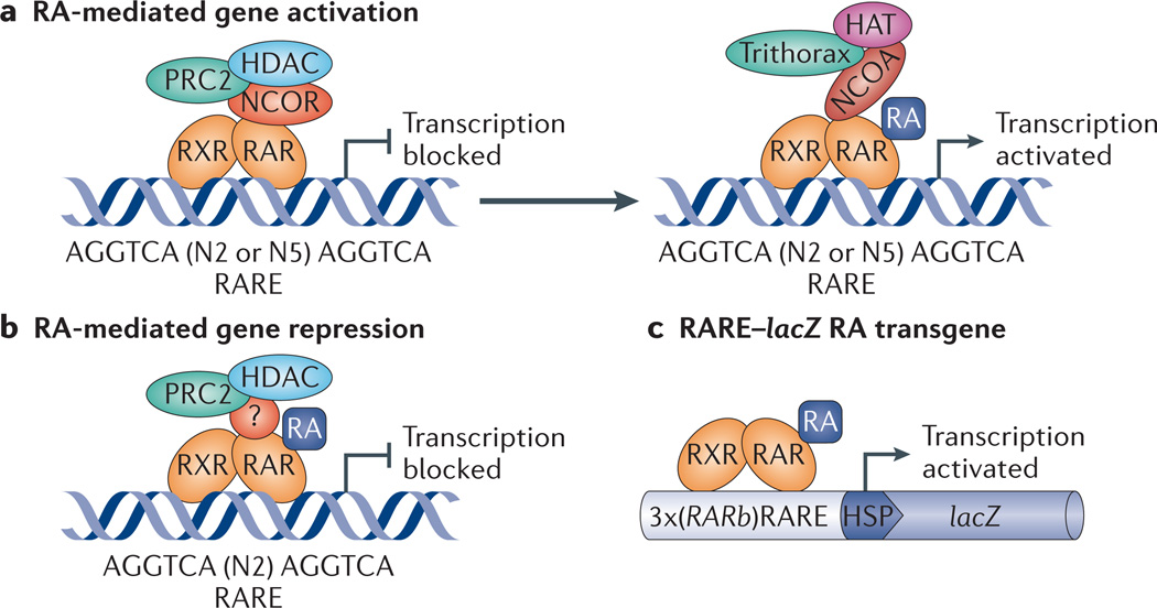
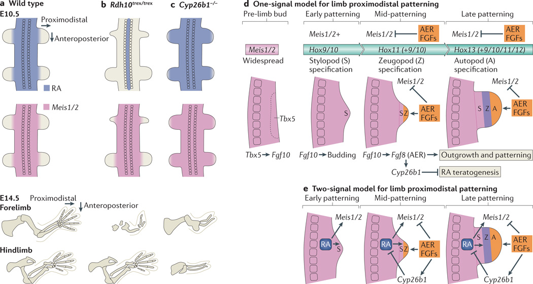
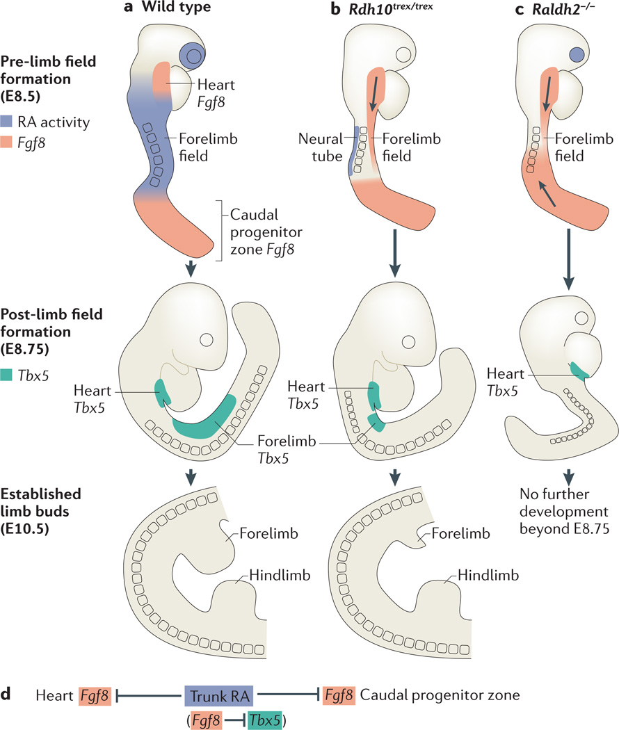
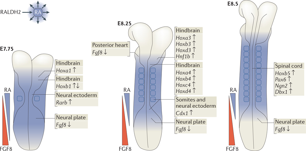
Retinoic acid (RA) signalling has a central role during vertebrate development. RA synthesized in specific locations regulates transcription by interacting with nuclear RA receptors (RARs) bound to RA response elements (RAREs) near target genes. RA was first implicated in signalling on the basis of its teratogenic effects on limb development. Genetic studies later revealed that endogenous RA promotes forelimb initiation by repressing fibroblast growth factor 8 (Fgf8). Insights into RA function in the limb serve as a paradigm for understanding how RA regulates other developmental processes. In vivo studies have identified RAREs that control repression of Fgf8 during body axis extension or activation of homeobox (Hox) genes and other key regulators during neuronal differentiation and organogenesis.


Figures

Figure 1
Figure 1. RA signalling mechanism
Retinoic acid (RA) binds to RA receptor (RAR) in an RAR–retinoid X receptor (RXR) heterodimer complex bound to RA response elements (RAREs) near target genes, resulting in control of transcription. a | For genes activated by RA, the absence of RA allows co-repressors of the nuclear receptor co-repressor (NCOR) family to bind to RAR and recruit repressive factors such as Polycomb repressive complex 2 (PRC2) and histone deacetylase (HDAC), whereas the presence of RA releases co-repressors and allows co-activators of the nuclear receptor co-activator (NCOA) family to bind to RAR and recruit activating factors such as Trithorax and histone acetylase (HAT). b | For genes repressed by RA (such as fibroblast growth factor 8 (Fgf8)), the presence of RA allows RAR to recruit PRC2 and HDAC (in this case the co-regulator, if any, is unknown, as indicated by ‘?’). c | The RARE–lacZ RA-reporter transgene, which is often used to monitor RA activity in vivo, consists of three tandem RAREs (from the Rarb gene) located upstream of a basal heat shock promoter (HSP) driving a lacZ gene cassette, which leads to expression of β-galactosidase.
Figure 2
Figure 2. Genetic studies indicate that limb patterning does not require RA signalling but does require FGF signalling and RA degradation
a | During limb-patterning stages, retinoic acid (RA) is prevented from entering the limb by the action of cytochrome P450 26B1 (CYP26B1). At the same time, expression of Meis1 and Meis2 (Meis1/2) marks the proximal limb. By embryonic day 14.5 (E14.5), all of the skeletal elements of the limbs have formed. b | In retinol dehydrogenase 10 (Rdh10) mouse mutants (Rdh10trex/trex), RA is limited to the neural tube and is missing from the limbs, whereas Meis1/2 expression in the limbs displays a normal distribution during patterning stages. Forelimbs are severely truncated from an early stage, which is indicative of their forelimb initiation defect, whereas hindlimbs have a normal complement of skeletal elements. c | In Cyp26b1–/– mouse mutants, RA degradation in the limbs is lost, and RA is detectable in more distal limb regions than wild-type mice during limb-patterning stages. Meis1/2 expression is also extended distally. At E14.5, all limb segments in both forelimbs and hindlimbs are significantly truncated. d | Based on mouse genetic studies, we propose a one-signal fibroblast growth factor (FGF)-driven progress-zone model for limb proximodistal patterning coupled with collinear homeobox (Hox) gene activation that does not require RA to specify proximal fate but requires RA degradation to prevent RA teratogenesis. Before limb budding, T-box 5 (Tbx5) activates Fgf10 in the limb field Fgf10 subsequently activates epithelial-to-mesenchymal transition and proliferation in the limb field, which leads to formation of the limb bud. Meis1/2 expression is present throughout the early bud and marks stylopod specification, which is dependent on Hox9 and Hox10 genes in the forelimb and Hox10 genes in the hindlimb. Subsequently, activation of Fgf8 by Fgf10 in the distal ectoderm leads to formation of the apical ectodermal ridge (AER) and continued distal FGF signalling that promotes outgrowth and activates Cyp26b1, which degrades RA to block RA-induced teratogenesis. AER FGF signals also repress Meis1/2 in the distal limb, which allows specification of a more distal zeugopod fate. This is dependent on Hox11 genes, which are activated later and more distally than Hox9 and Hox10 genes in an autonomous manner. Later activation of Hox13 and Hox12 genes at the distal extremity is required for autopod specification. e | Chick studies support a two-signal model for control of limb proximodistal patterning in which RA is required to establish proximal fate, whereas an opposing FGF signal is required for distal fate. RA generated in the trunk diffuses into the proximal limb to stimulate Meis1/2 expression, and FGF generated distally in the AER then represses Meis1/2 and induces Cyp26b1 to degrade RA distally to limit Meis1/2 expression to the proximal limb.
Figure 3
Figure 3. RA– Fgf8 antagonism regulates the initiation of forelimb budding
a | Before forelimb field formation, wild-type mouse embryos have two domains of fibroblast growth factor 8 (Fgf8) expression (red) in the heart and caudal progenitor zone, flanking regions of retinoic acid (RA) signalling in the trunk (blue). T-box 5|( Tbx5) expression (green) in the lateral plate mesoderm marks the established forelimb field. By embryonic day 10.5 (E10.5), forelimb and hindlimb buds are clearly established. b | In retinol dehydrogenase 10 (Rdh10)-mutant mice (Rdh10trex/trex), loss of mesodermal RA activity is coupled with a posterior extension of cardiac Fgf8 expression into the presumptive forelimb field. Limb Tbx5 expression is subsequently delayed, and the region of Tbx5 expression is significantly shortened along the anteroposterior axis. At E10.5, forelimbs are hypoplastic, whereas hindlimbs are unaffected. c | In retinaldehyde dehydrogenase 2 (Raldh2)-mutant mice, a lack of RA activity in the trunk (both mesoderm and neuroectoderm) is coupled with two fronts of ectopic Fgf8 expression into the presumptive forelimb field, from both the heart and the caudal progenitor zone. Limb Tbx5 expression is subsequently prevented. Raldh2−/− mutants stop developing at around E8.75. d | RA–Fgf8 antagonism is tightly associated with limb Tbx5 activation.
Figure 4
Figure 4. Direct RA target genes that are required for normal body axis extension, anteroposterior patterning, neurogenesis and somitogenesis
Retinoic acid (RA) generated by retinaldehyde dehydrogenase 2(RALDH2) in trunk mesoderm diffuses to nearby tissues to control several aspects of early mouse development. An overarching theme during these early stages is repression of caudal fibroblast growth factor 8 (Fgf8) by RA (depicted by opposing gradients of RA and FGF8) to allow normal body axis extension. In addition, RA activates homeobox (Hox) genes (and other genes) that are required for anteroposterior patterning of the trunk, neurogenesis and somitogenesis. Target genes are indicated with up-arrows for RA activation and down-arrows for RA repression. Cdx1, caudal-type homeobox 1; Dbx1, developing brain homeobox 1; Hnf1b, HNF1 homeobox B; Ngn2, neurogenin 2; Pax6, paired box 6; Rarb, retinoic acid receptor-β.
Figure 5
Figure 5. Diverse roles of RA in regulating mouse organogenesis
a | Eye: retinoic acid (RA) generated by retinaldehyde dehydrogenase 1 (RALDH1) and RALDH3 in the retina activates paired-like homeodomain transcription factor 2 (Pitx2) in perioptic mesenchyme, resulting in activation of dickkopf homologue 2 (Dkk2), which downregulates WNT signalling. b | In the late forebrain basal ganglia, RA generated by RALDH3 in the lateral ganglionic eminence and septum promotes γ-aminobutyric acid (GABA)-ergic differentiation by an unknown mechanism and stimulates dopamine signalling by activating dopamine receptor D2 (Drd2) in the nucleus accumbens within the striatum. c | In the heart and liver, RA generated by RALDH2 in the liver mesothelium activates erythropoietin (Epo) in fetal liver. Epo locally stimulates erythropoiesis and, via transport of EPO in the circulatory system, stimulates myocardial proliferation through EPO-mediated upregulation of insulin-like growth factor 2 (Igf2) in epicardium. d | In the postnatal testis, RA generated by RALDH2 in spermatocytes activates Stra8 (stimulated by retinoic acid gene 8) for the induction of meiosis.

References

    1. Wolbach SB, Howe PR. Tissue changes following deprivation of fat-soluble A vitamin. J. Exp. Med. 1925;42:753–777. - PMC - PubMed
    1. Frazier CN, Hu CK. Cutaneous lesions associated with a deficiency in vitamin A in man. Arch. Intern. Med. 1931;48:507–514.
    1. Wilson JG, Roth CB, Warkany J. An analysis of the syndrome of malformations induced by maternal vitamin A deficiency. Effects of restoration of vitamin A at various times during gestation. Amer. J. Anat. 1953;92:189–217. - PubMed
    1. Clagett-Dame M, DeLuca HF. The role of vitamin A in mammalian reproduction and embryonic development. Annu. Rev. Nutr. 2002;22:347–381. - PubMed
    1. Dowling JE, Wald G. Vitamin A deficiency and nigh blindness. Proc. Natl Acad. Sci. USA. 1958;44:648–661. - PMC - PubMed

Publication types

MeSH terms