. 2015 Jan 27;9(1):809-16.
doi: 10.1021/nn5063497. Epub 2015 Jan 8.

Combing of genomic DNA from droplets containing picograms of material

Affiliations

Combing of genomic DNA from droplets containing picograms of material

Jochem Deen et al. ACS Nano. .

Abstract

Deposition of linear DNA molecules is a critical step in many single-molecule genomic approaches including DNA mapping, fiber-FISH, and several emerging sequencing technologies. In the ideal situation, the DNA that is deposited for these experiments is absolutely linear and uniformly stretched, thereby enabling accurate distance measurements. However, this is rarely the case, and furthermore, current approaches for the capture and linearization of DNA on a surface tend to require complex surface preparation and large amounts of starting material to achieve genomic-scale mapping. This makes them technically demanding and prevents their application in emerging fields of genomics, such as single-cell based analyses. Here we describe a simple and extremely efficient approach to the deposition and linearization of genomic DNA molecules. We employ droplets containing as little as tens of picograms of material and simply drag them, using a pipet tip, over a polymer-coated coverslip. In this report we highlight one particular polymer, Zeonex, which is remarkably efficient at capturing DNA. We characterize the method of DNA capture on the Zeonex surface and find that the use of droplets greatly facilitates the efficient deposition of DNA. This is the result of a circulating flow in the droplet that maintains a high DNA concentration at the interface of the surface/solution. Overall, our approach provides an accessible route to the study of genomic structural variation from samples containing no more than a handful of cells.

Keywords: DNA deposition; DNA mapping; coffee ring effect; fiber-FISH; molecular combing; rolling droplet; single-molecule imaging.


Figures

Figure 1
Figure 1
DNA deposition on Zeonex from an evaporating droplet. DNA forms a stable attachment to the surface (yellow image), and the vast majority of these molecules are stretched (blue) as the contact line moves over them as shown in the merged image. DNA molecules are stained with 50 nM SYBR Gold. Scale bar is 20 μm in length.
Figure 2
Figure 2
Schematic representation of our rolling droplet experiment and microscope images taken of DNA deposition at the edge of droplets translated across the three different surfaces. Deposition is far more efficient on the Zeonex surface than other substrates. The surface is translated below the droplet with a speed of 1.5 mm/min. Scale bars are 10 μm in length. DNA is stained with 50 nM SYBR Gold dye (a nonspecific, groove-binding fluorophore).
Figure 3
Figure 3
Long-exposure confocal microscopy images using fluorescent tracer beads to depict the flow in a microliter droplet as it is translated across a Zeonex-coated substrate. The six microscopy images show the movement of the tracer beads at the front (close to the pipet tip) and the back of the droplet, at different depths for a 5 s camera exposure. Note that there are approximately five times more particles at the surface interface compared with the bulk solution. Scale bar is 100 μm. The right panel shows the modeled flow profile in the droplet, in the reference frame of the droplet, derived using the microscopy data. Arrows are vectors indicating the velocity of the flow around the droplet. The circulating flow in the droplet results in a continuous supply of DNA molecules to the surface, where they can subsequently bind the surface and be stretched.
Figure 4
Figure 4
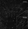
Fluorescence microscopy images (using an inverted look-up table, where black depicts high photon counts) of YOYO-1-stained human genomic DNA (extracted from HeLa cells) deposited from a 1 μL droplet containing (a, b) approximately 170 pg of DNA or (c) 40 pg of DNA. All scale bars are 200 kilobase pairs in length (108 μm). DNA is deposited and linearized perpendicular to the edge of the rolling droplet resulting in a characteristic deposition pattern.
Figure 5
Figure 5
A total of 1225 tiled fluorescence microscopy images of a sample of E. coli genomic DNA labeled with Atto647N at sites reading 5′-TCGA-3′ using the M.TaqI DNA methyltransferase enzyme in order to direct labeling. The image contains approximately 500 megabases of genomic material and took of the order of 10 min to acquire.

References

    1. Neely R. K.; Deen J.; Hofkens J. Optical Mapping of DNA: Single-Molecule-Based Methods for Mapping Genomes. Biopolymers 2011, 95, 298–311. - PubMed
    1. Samad A. H.; Cai W. W.; Hu X.; Irvin B.; Jing J.; Reed J.; Meng X.; Huang J.; Huff E.; Porter B.; et al. Mapping the Genome One Molecule at a Time - Optical Mapping. Nature 1995, 378, 516–517. - PubMed
    1. Teague B.; Waterman M. S.; Goldstein S.; Potamousis K.; Zhou S.; Reslewic S.; Sarkar D.; Valouev A.; Churas C.; Kidd J. M.; et al. High-Resolution Human Genome Structure by Single-Molecule Analysis. Proc. Natl. Acad. Sci. U.S.A. 2010, 107, 10848–10853. - PMC - PubMed
    1. Kim S.; Gottfried A.; Lin R. R.; Dertinger T.; Kim A. S.; Chung S.; Colyer R. A.; Weinhold E.; Weiss S.; Ebenstein Y. Enzymatically Incorporated Genomic Tags for Optical Mapping of DNA-Binding Proteins. Angew. Chem., Int. Ed. 2012, 51, 3578–3581. - PMC - PubMed
    1. Michalet X.; Ekong R.; Fougerousse F.; Rousseaux S.; Schurra C.; Hornigold N.; Slegtenhorst M. v.; Wolfe J.; Povey S.; Beckmann J. S.; et al. Dynamic Molecular Combing: Stretching the Whole Human Genome for High-Resolution Studies. Science 1997, 277, 1518–1523. - PubMed

Publication types