. 2015 May;180(2):305-15.
doi: 10.1111/cei.12579.

CXCL10 induces the recruitment of monocyte-derived macrophages into kidney, which aggravate puromycin aminonucleoside nephrosis

Affiliations

CXCL10 induces the recruitment of monocyte-derived macrophages into kidney, which aggravate puromycin aminonucleoside nephrosis

D Petrovic-Djergovic et al. Clin Exp Immunol. 2015 May.

Abstract

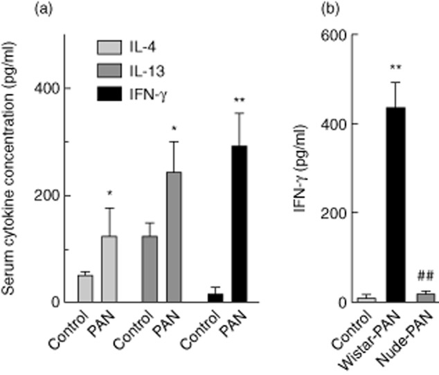
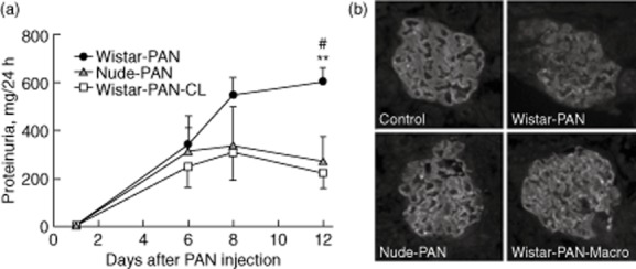
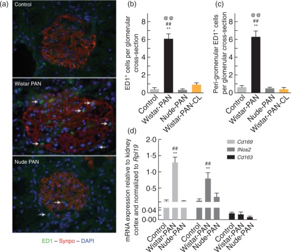
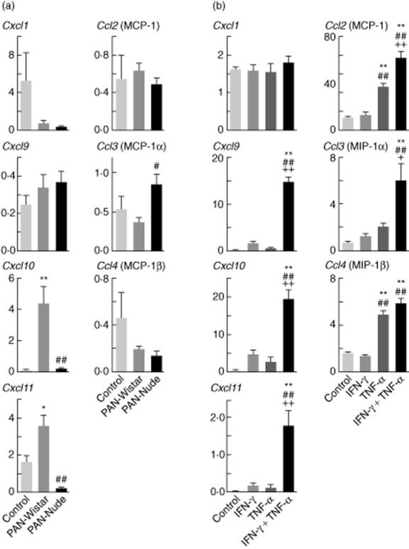
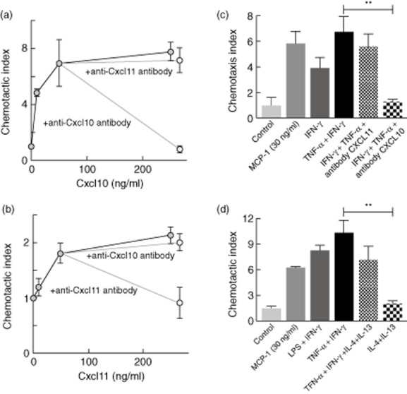
The mechanism responsible for trafficking of monocyte-derived macrophages into kidney in the puromycin aminonucleoside model of nephrotic syndrome in rats (PAN-NS), and the significance of this infiltration, remain largely unknown. CXCL10, a chemokine secreted in many T helper type 1 (Th1) inflammatory diseases, exhibits important roles in trafficking of monocytes and activated T cells. We hypothesized that induction of circulating interferon (IFN)-γ and glomerular tumour necrosis factor (TNF)-α during PAN-NS would stimulate the release of CXCL10 by podocytes, leading to infiltration of activated immune cells and greater glomerular injury. We found that serum IFN-γ, glomerular Cxcl10 mRNA and intra- and peri-glomerular macrophage infiltration were induced strongly during the late acute phase of PAN-NS in Wistar rats, but not in nude (Foxn1(rnu/rnu) ) rats lacking functional effector T lymphocytes. Wistar rats also developed significantly greater proteinuria than nude rats, which could be abolished by macrophage depletion. Stimulation of cultured podocytes with both IFN-γ and TNF-α markedly induced the expression of Cxcl10 mRNA and CXCL10 secretion. Together, these data support our hypothesis that increased circulating IFN-γ and glomerular TNF-α induce synergistically the production and secretion of CXCL10 by podocytes, attracting activated macrophages into kidney tissue. The study also suggests that IFN-γ, secreted from Th1 lymphocytes, may prime proinflammatory macrophages that consequently aggravate renal injury.

Keywords: CXCL10; chemokines; kidney injury; macrophages; nephrotic syndrome.


Figures

Fig 1
Fig 1
Serum interferon (IFN)-γ is induced at the peak of proteinuria induced by puromycin aminonucleoside (PAN) in Wistar but not nude (Foxn1rnu/rnu) rats. (a) Serum cytokine concentrations in blood samples taken from Wistar rats at 12 days after injection with vehicle alone (n = 4) or PAN (n = 8). (b) Serum IFN-γ concentrations in blood samples taken from Wistar or nude rats at 12 days after injection with vehicle alone (n = 4) or PAN (n = 8). Results are expressed as means ± standard error; *P < 0·05; **P < 0·01 compared with vehicle alone by unpaired, two-tailed t-test.
Fig 2
Fig 2
Proteinuria and podocyte injury in late stages of acute puromycin aminonucleoside (PAN) nephrosis are greater in Wistar rats than in nude rats, or in Wistar rats depleted of macrophages. (a) Plot of protein concentrations in 24-h urine samples taken at various times after PAN injection from Wistar (Wistar-PAN, n = 8) nude rats (nude-PAN, n = 4) and from Wistar rats whose macrophages were depleted by three intraperitoneal (i.p.) injections of clodronate liposomes at 4, 7 and 10 days after PAN injection (Wistar-PAN-CL, n = 4). Results are expressed as means ± standard error; **P < 0·01 compared with nude-PAN and #P < 0·05 compared with Wistar-PAN-CL by analysis of variance (anova) followed by Bonferroni's post-test. (b) Micrographs visualizing anti-synaptopodin antibody labelling of representative glomeruli in frozen sections from the same animals used in the experiment shown in (a), along with sham-injected control animals (control). Frozen sections were obtained from animals killked 12 days after PAN injection.
Fig 3
Fig 3
Activated (ED1+) macrophages accumulate in the glomerulus and peri-glomerular areas of Wistar rats [Wistar-puromycin aminonucleoside (PAN)], but not of nude rats (nude-PAN) or macrophage-depleted Wistar rats (Wistar-PAN-CL), during the late stage of acute PAN nephrosis. (a) Representative micrographs of glomeruli labelled with antibodies directed against ED1 (green), synaptopodin (red) and 4′,6-diamidino-2-phenylindole (DAPI) (blue) from Wistar rats injected with vehicle alone (control) or PAN (Wistar-PAN), or from nude rats injected with PAN (nude-PAN). The arrows show positions of glomerular and peri-glomerular activated (ED1+) macrophages. Results in studies of macrophage-depleted Wistar rats were similar to those shown for nude rats (not shown). Quantitative results of immunofluorescence analyses are shown in plots of intra- and peri-glomerular macrophage counts from these animals (b), including macrophage-depleted Wistar rats (Wistar-PAN-CL). Results are expressed as means ± standard error (s.e.), n = 4/group; **P < 0·01 compared with vehicle alone, ##P < 0·01 compared to nude-PAN, @@P < 0·01 compared to Wistar-PAN-CL. Shown in (d) are plots of the mRNA expression of markers of proinflammatory activated macrophages (Cd169, Nos2), and of alternatively activated macrophages (Cd163). Results are expressed as means ± s.e., n = 4/group; **P < 0·01 compared to vehicle alone and ##P < 0·01 compared to nude-PAN.
Fig 4
Fig 4
The expression of Cxcl10 and Cxcl11 is induced during late acute puromycin aminonucleoside (PAN) nephrosis in Wistar but not nude rats, and synergistically by interferon (IFN)-γ and tumour necrosis factor (TNF)-α in cultured human podocytes. (a) Plots of mRNA expression of various chemokines, relative to normal kidney cortex and normalized to Rpl19. The mRNA expression of genes encoding the C-X-C chemokines (Cxcl1, Cxcl9, Cxcl10, Cxcl11) as well as C-C chemokines (Ccl2, Ccl3, Ccl4) was evaluated in glomeruli isolated 12 days after Wistar or nude rats were injected with PAN (PAN-Wistar, PAN-nude), or with vehicle alone (control). Results are expressed as means ± standard error (s.e.), n = 4/group; *P < 0·05 compared with vehicle alone; **P < 0·01 or ##P < 0·01 compared with Wistar-PAN and vehicle alone. (b) Normalized expression of similar set of C-X-C and C-C chemokine genes as in (a). The mRNA expression was evaluated in cultured human podocytes after 4 h of treatment with IFN-γ, TNF-α or both cytokines together (each at 10 ng/ml). Results are expressed as means ± s.e., n = 6/group, **P < 0·01 compared with control, ##P < 0·01 compared with IFN-γ or ++P < 0·01 compared with TNF-α.
Fig 5
Fig 5
CXCL10 produced by cultured human podocytes in response to interferon (IFN)-γ and tumour necrosis factor (TNF)-α induces monocyte migration. The chemotactic index was calculated as the fold increase, compared to sham-treated controls, in the number of THP-1 monocytes migrating across a 5-μm-pore membrane in response to chemoattractants. (a) Migration after incubation over wells containing various concentrations of recombinant CXCL10, or with 250 ng of CXCL10/ml preincubated with neutralizing antibodies against CXCL10 or CXCL11. (b) Migration after incubation over wells containing various concentrations of recombinant CXCL11, or with 250 ng of CXCL11/ml preincubated with neutralizing antibodies against CXCL10 or CXCL11. (c) Migration after incubation over wells containing culture supernatants from human podocytes collected 24 h after a 6 h treatment with IFN-γ, or TNF-α + IFN-γ. (d) Migration after incubation over wells containing culture supernatants from human podocytes collected 24 h after a 6 h treatment with either type 1 stimulation [lipopolysaccharide (LPS) + IFN-γ, or TNF-α + IFN-γ] or type 1 + type 2 stimulation (TNF-α + IFN-γ + IL-4 + IL-13) or type 2 cytokines only (IL-4 + IL-13). Control supernatants are from untreated podocyte cultures. The chemoattractant CCL2 [monocyte chemoattractant protein-1 (MCP-1)] was used as positive control. Results are expressed as mean ± standard error, n = 4/group; **P < 0·01 compared to control.

References

    1. Anderson S, Diamond JR, Karnovsky MJ, Brenner BM. Mechanisms underlying transition from acute glomerular injury to late glomerular sclerosis in a rat model of nephrotic syndrome. J Clin Invest. 1988;82:1757–1768. - PMC - PubMed
    1. Hoyer JR, Ratte J, Potter AH, Michael AF. Transfer of aminonucleoside nephrosis by renal transplantation. J Clin Invest. 1972;51:2777–2780. - PMC - PubMed
    1. Schreiner GF, Cotran RS, Unanue ER. Modulation of Ia and leukocyte common antigen expression in rat glomeruli during the course of glomerulonephritis and aminonucleoside nephrosis. Lab Invest. 1984;51:524–533. - PubMed
    1. Ricardo SD, van Goor H, Eddy AA. Macrophage diversity in renal injury and repair. J Clin Invest. 2008;118:3522–3530. - PMC - PubMed
    1. Diamond JR, Pesek I, Ruggieri S, Karnovsky MJ. Essential fatty acid deficiency during acute puromycin nephrosis ameliorates late renal injury. Am J Physiol. 1989;257:F798–807. - PubMed

Publication types

MeSH terms