. 2015 Jan 1;29(1):39-47.
doi: 10.1101/gad.246173.114.

Stably paused genes revealed through inhibition of transcription initiation by the TFIIH inhibitor triptolide

Affiliations

Stably paused genes revealed through inhibition of transcription initiation by the TFIIH inhibitor triptolide

Fei Chen et al. Genes Dev. .

Abstract

Transcription by RNA polymerase II (Pol II) in metazoans is regulated in several steps, including preinitiation complex (PIC) formation, initiation, Pol II escape, productive elongation, cotranscriptional RNA processing, and termination. Genome-wide studies have demonstrated that the phenomenon of promoter-bound Pol II pausing is widespread, especially for genes involved in developmental and stimulus-responsive pathways. However, a mechanistic understanding of the paused Pol II state at promoters is limited. For example, at a global level, it is unclear to what extent the engaged paused Pol II is stably tethered to the promoter or undergoes rapid cycles of initiation and termination. Here we used the small molecule triptolide (TPL), an XPB/TFIIH inhibitor, to block transcriptional initiation and then measured Pol II occupancy by chromatin immunoprecipitation (ChIP) followed by next-generation sequencing (ChIP-seq). This inhibition of initiation enabled us to investigate different states of paused Pol II. Specifically, our global analysis revealed that most genes with paused Pol II, as defined by a pausing index, show significant clearance of Pol II during the period of TPL treatment. Our study further identified a group of genes with unexpectedly stably paused Pol II, with unchanged Pol II occupancy even after 1 h of inhibition of initiation. This group of genes constitutes a small portion of all paused genes defined by the conventional criterion of pausing index. These findings could pave the way for evaluating the contribution of different elongation/pausing factors on different states of Pol II pausing in developmental and other stimulus-responsive pathways.

Keywords: RNA polymerase II; chromatin; transcription.


Figures

Figure 1.
Figure 1.
The effect of TPL treatment on Pol II levels and transcriptional activity. (A) Western blot analysis for Pol II in untreated (0 min) and TPL-treated (125 nM) HCT116 cells. Total Pol II (N20), Ser5 phosphorylated Pol II (H14), Ser2 phosphorylated Pol II (H5), and hypophosphorylated Pol II (8WG16) antibodies were used to determine Pol II levels. (BD) qRT-PCR analysis of expression levels of serum-inducible genes EGR1 (B), FOS (C), and JUNB (D) in HCT116 cells. Cells were starved for 48 h before the addition of serum for 30 min. TPL was added for the indicated time points, except vehicle (DMSO)-treated cells (0 min TPL; blue bar) and starved cells (0 min TPL; gray bar).
Figure 2.
Figure 2.
TPL treatment alters Pol II occupancy. (A) Metagene plots of Pol II occupancy for all genes with (red) or without (black) 60 min of TPL treatment. HCT116 cells were starved for 48 h before the addition of serum for 24 min and ChIP-seq analysis for RNA Pol II (N20 antibody). Global loss of Pol II on both promoters and gene bodies was observed as the result of TPL treatment. The shaded region indicates the 95% confidence interval of the difference between two medians. (B) Metagene plot of the regions −100 base pairs (bp) to +200 bp around the TSS, plotted as the percentage of maximum for each condition. Dashed lines indicate the peak summit. In TPL-treated cells, Pol II peaks more toward the 5′ of the gene than in the DMSO-treated cells. (C) Metagene analysis as in A, but with genes divided into serum-induced (top panel) and uninduced (bottom panel), is shown. The insets show the 5′ shift of Pol II at promoters for each gene class. (D) Genome browser track examples of Pol II occupancy at promoter regions for FOS, JUNB, and HIST1H2A with (red) or without (black) TPL treatment. Dashed lines indicate the annotated TSS for each gene. (E) Model for the observed shift in Pol II occupancy observed after TPL treatment. (Top panel) The promoter-bound Pol II peak is composed of nonengaged Pol II and engaged paused Pol II. The transition from the nonengaged Pol II to the engaged Pol II, transcribing ∼20–60 nt, requires the helicase/translocase activity of XPB/TFIIH. (Middle panel) Upon receipt of cellular signals (e.g., +serum), engaged paused Pol II is phosphorylated by P-TEFb/SEC, switches to productively elongating Pol II, and continues into gene bodies. Under normal conditions, release from the paused state allows new polymerases to enter the engaged state in a TFIIH-dependent manner. (Bottom panel) Upon treatment of TPL, the already engaged but transiently pausing Pol II can still escape into gene bodies to complete transcription, but the transition from nonengaged Pol II to engaged Pol II is blocked, which leads to the observed decrease and 5′ shift in promoter-proximal Pol II occupancy.
Figure 3.
Figure 3.
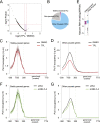
Different degrees of pausing. (A–I) Genome browser track examples showing the contrast between stably paused Pol II and other forms of paused Pol II. (A) Schematic presentation showing the experimental design. Serum starvation began 48 h prior to harvesting and fixation of the cells for ChIP-seq; cells were treated with TPL or the vehicle DMSO for 1 h prior to fixation. For serum-activated cells, serum was added 8 min prior to fixation. (B,D,F,H) Genome browser track examples of Pol II occupancies in the serum-starved condition and with treatment with TPL or DMSO. (C,E,G,I) Genome browser track examples of Pol II occupancies in the serum-activated condition and with treatment with TPL or DMSO.
Figure 4.
Figure 4.
Global identification and characterization of stably paused genes. (A) Volcano plot representing the criteria for the classification of Pol II on stably paused genes. Stably paused genes, highlighted in pink, are the subset of paused genes with a log2 fold change in Pol II occupancy during 1 h of TPL treatment of <0.15 and a P-value for differential occupancy of >0.05 (−log10 P-value < 1.3). (B) Pie chart representing the percentage of stably paused genes, other forms of paused genes, and nonpaused genes. (C,D) Metagene analysis of Pol II occupancy for stably paused genes with (red) or without (black) TPL treatment. (E) Box plot analysis of nascent RNA levels for stably paused genes and other forms of paused genes, confirming that stably paused genes release less productively elongating Pol II than the majority of genes exhibiting paused Pol II. (F,G) Metagene analysis of Pol II occupancy for stably paused genes and other forms of paused genes targeted by a scrambled shRNA (shScr; black) or NELF-A shRNA (shNELF-A; lime).

References

    1. Adelman K, Lis JT. 2012. Promoter-proximal pausing of RNA polymerase II: emerging roles in metazoans. Nat Rev Genet 13: 720–731. - PMC - PubMed
    1. Boehm AK, Saunders A, Werner J, Lis JT. 2003. Transcription factor and polymerase recruitment, modification, and movement on dhsp70 in vivo in the minutes following heat shock. Mol Cell Biol 23: 7628–7637. - PMC - PubMed
    1. Brannan K, Kim H, Erickson B, Glover-Cutter K, Kim S, Fong N, Kiemele L, Hansen K, Davis R, Lykke-Andersen J, et al. . 2012. mRNA decapping factors and the exonuclease Xrn2 function in widespread premature termination of RNA polymerase II transcription. Mol Cell 46: 311–324. - PMC - PubMed
    1. Buckley MS, Kwak H, Zipfel WR, Lis JT. 2014. Kinetics of promoter Pol II on Hsp70 reveal stable pausing and key insights into its regulation. Genes Dev 28: 14–19. - PMC - PubMed
    1. Cheng B, Li T, Rahl PB, Adamson TE, Loudas NB, Guo J, Varzavand K, Cooper JJ, Hu X, Gnatt A, et al. . 2012. Functional association of Gdown1 with RNA polymerase II poised on human genes. Mol Cell 45: 38–50. - PMC - PubMed

Publication types