. 2015 Jan 1;29(1):94-107.
doi: 10.1101/gad.253070.114.

The G-patch protein Spp2 couples the spliceosome-stimulated ATPase activity of the DEAH-box protein Prp2 to catalytic activation of the spliceosome

Affiliations

The G-patch protein Spp2 couples the spliceosome-stimulated ATPase activity of the DEAH-box protein Prp2 to catalytic activation of the spliceosome

Zbigniew Warkocki et al. Genes Dev. .

Abstract

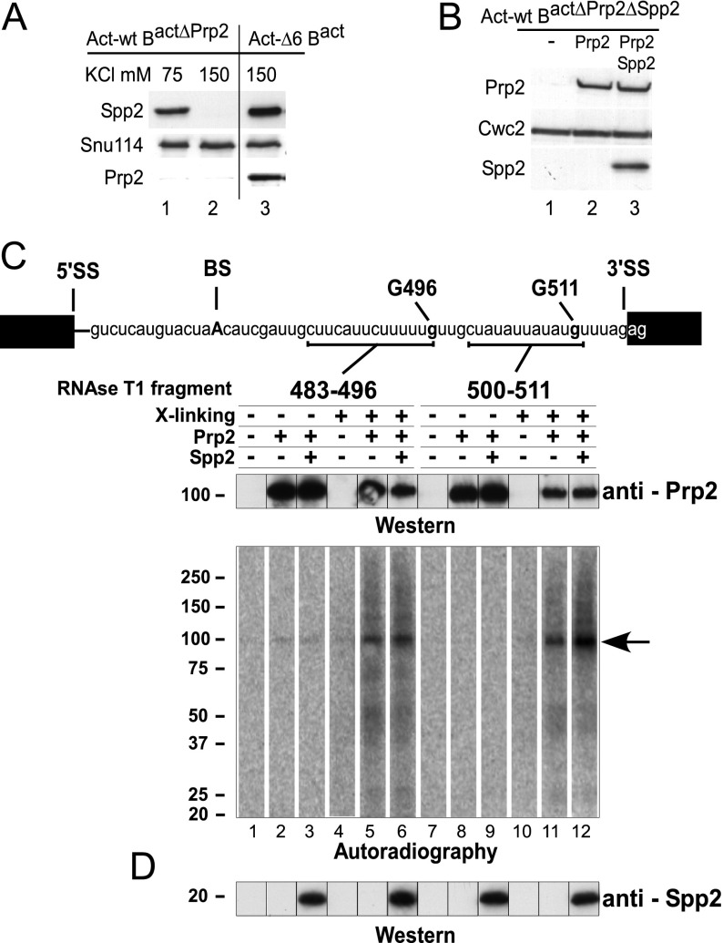
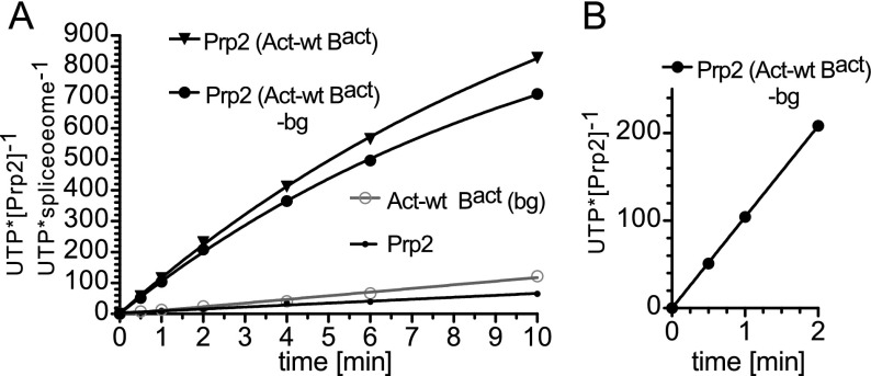
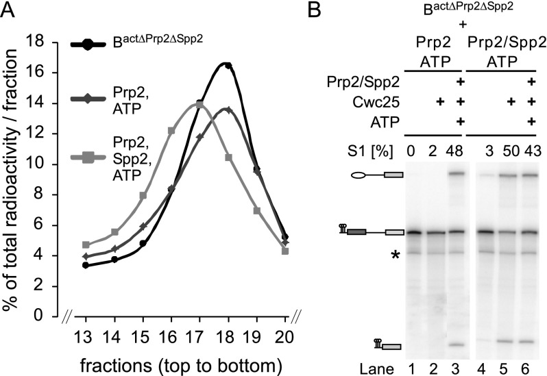
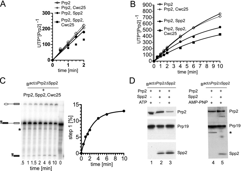
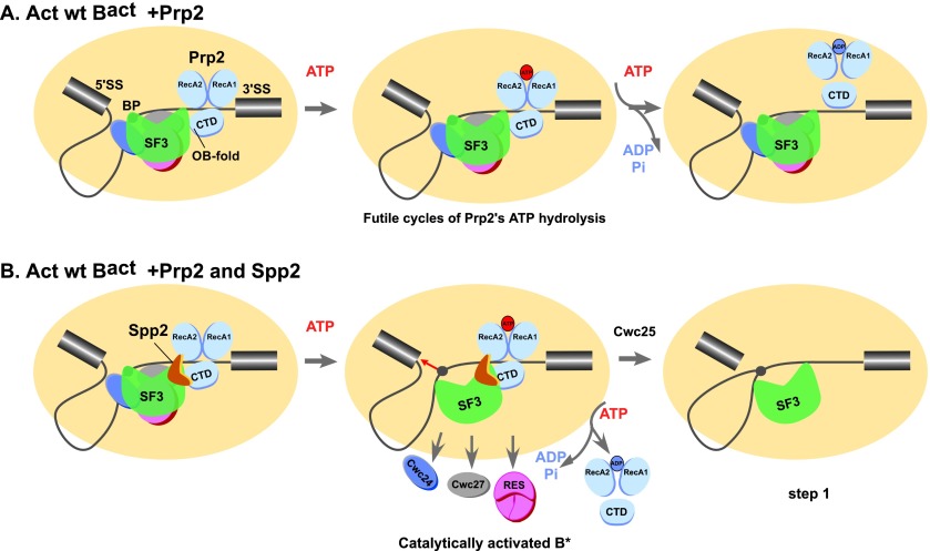
Structural rearrangement of the activated spliceosome (B(act)) to yield a catalytically active complex (B*) is mediated by the DEAH-box NTPase Prp2 in cooperation with the G-patch protein Spp2. However, how the energy of ATP hydrolysis by Prp2 is coupled to mechanical work and what role Spp2 plays in this process are unclear. Using a purified splicing system, we demonstrate that Spp2 is not required to recruit Prp2 to its bona fide binding site in the B(act) spliceosome. In the absence of Spp2, the B(act) spliceosome efficiently triggers Prp2's NTPase activity, but NTP hydrolysis is not coupled to ribonucleoprotein (RNP) rearrangements leading to catalytic activation of the spliceosome. Transformation of the B(act) to the B* spliceosome occurs only when Spp2 is present and is accompanied by dissociation of Prp2 and a reduction in its NTPase activity. In the absence of spliceosomes, Spp2 enhances Prp2's RNA-dependent ATPase activity without affecting its RNA affinity. Our data suggest that Spp2 plays a major role in coupling Prp2's ATPase activity to remodeling of the spliceosome into a catalytically active machine.

Keywords: ATP hydrolysis; DEAH-box helicase; G-patch protein; Prp2; Spp2; spliceosome activation.


Figures

Figure 1.
Figure 1.
Prp2 and Spp2 bind to the spliceosome independently. (A) Western blot analysis of Spp2 association with the spliceosome. Spliceosomes were assembled on wild-type (lanes 1,2) or truncated (ActΔ6) actin pre-mRNAs (which leads to stalling of the spliceosome assembly at the Bact stage) (lane 3) in heat-treated prp2-1 extract (lanes 1,2) or wild-type extract (lane 3). Spliceosomes were affinity-purified in parallel in the presence of 75 mM (lane 1) or 150 mM (lanes 2,3) KCl. Immunoblotting was performed with rabbit polyclonal antibodies against GST-Spp2, Prp2, and Snu114 as indicated. (B) Prp2 and Spp2 association with the spliceosome at 150 mM KCl. The Bact ΔPrp2 ΔSpp2 spliceosomes were incubated with buffer (lane 1), Prp2 (lane 2), or Prp2 and Spp2 (lane 3). Unbound proteins were removed by washing with buffer containing 150 mM KCl, and spliceosomes were fractionated by glycerol gradient centrifugation at 150 mM KCl. Immunoblotting was performed with rabbit polyclonal antibodies against Prp2, Spp2, and Cwc2. (C) Cross-linking of Prp2 to pre-mRNA in purified spliceosomes. Two distinct site-specifically labeled pre-mRNAs were created, each carrying a single 32P-labeled phosphate 5′ at nucleotide G 496 or G 511. The theoretical RNA fragments remaining after digestion with RNase T1 are indicated by a bar below the sequence. Bact ΔPrp2 ΔSpp2 spliceosomes were assembled on site-specifically modified actin pre-mRNAs and purified at 150 mM KCl. After incubating with Prp2 or Prp2 plus Spp2, the complexes were UV cross-linked and then digested with RNase T1. Proteins cross-linked to 32P-labeled RNA were precipitated with antibodies against Prp2 and analyzed by SDS PAGE. Cross-linked 32P-labeled proteins were visualized by Western blot analysis (top panel) and autoradiography (bottom panel) as described above. The arrow indicates the 32P-labeled RNA cross-linked to Prp2. (D) Bact spliceosomes before cross-linking were probed with antibodies against Spp2.
Figure 2.
Figure 2.
The spliceosome is a potent stimulator of Prp2’s NTPase activity. (A) UTP hydrolysis was investigated by TLC and quantified by a PhosphorImager. UTP hydrolysis by the purified Bact ΔPrp2 ΔSpp2 spliceosomes in either the absence [Act-wt Bact (bg)] or presence (Prp2 Act-wt Bact) of Prp2 or by Prp2 alone (Prp2) was determined. The amount of UTP hydrolyzed by Prp2 in spliceosomes (“UTP•spliceosome−1”) and hydrolyzed by Prp2 in the absence of the spliceosome (Prp2) (“UTP•[Prp2]−1”) is plotted as a function of time. Prp2 Act-wt Bact (−bg) was generated by subtracting the spliceosome alone [Act-wt Bact (bg)] values from those obtained with Prp2 Act-wt Bact. (B) Initial rate of UTP hydrolysis by Prp2 in spliceosomes within the first 2 min of the time course. Data points were analyzed by linear regression to derive the initial rate of Prp2-catalyzed UTP hydrolysis in the context of the spliceosome.
Figure 3.
Figure 3.
Spp2 is required for Prp2-catalyzed NTP-dependent remodeling of the Bact spliceosome. (A) Glycerol gradient sedimentation profiles of Act-wt Bact ΔPrp2 ΔSpp2 spliceosomes incubated with buffer alone (black) or in the presence of Prp2 and ATP in either the absence (dark gray) or presence (light gray) of Spp2. Radioactivity contained in each fraction was determined by Cherenkov counting and calculated as the percentage of total radioactivity in one gradient. The percentage of total radioactivity present in each gradient fraction is plotted. Ten percent to 30% (v/v) glycerol gradients containing 75 mM KCl were loaded with 400-µL samples and centrifuged at 60,000 rpm for 2 h in a TH660 rotor (Sorvall). (B) The Bact ΔPrp2 ΔSpp2 spliceosomes complemented with Prp2/ATP or Prp2/Spp2/ATP were recovered from the peak fractions of the glycerol gradients shown in A and then incubated for 1 h at 23°C under reconstitution conditions with buffer (lanes 1,4), Cwc25 (lanes 2,5), or Prp2, Spp2, Cwc25, and ATP (lanes 3,6, positive controls). Thus, in lanes 46, spliceosomes that had been catalytically activated during the preincubation step were used. The formation of step 1 splicing products was monitored by 8% denaturing RNA PAGE and quantified by a PhosphorImager. The percentage of step 1 (S1) products (compared with the total RNA signal in a lane) is indicated above each lane. RNA species are indicated at the left (from the top): lariat–intron–3′ exon, pre-mRNA, uncharacterized RNA species, and 5′ exon.
Figure 4.
Figure 4.
UTP hydrolysis by Prp2 in the spliceosome is reduced after B* formation. (A) UTP hydrolysis was monitored by TLC and quantified by a PhosphorImager. Bact ΔPrp2 ΔSpp2 spliceosomes were incubated with Prp2 at an ∼1:1 ratio in the absence or presence of a 3–5 molar excess (140–150 nM) of Spp2, Spp2 and Cwc25, or Cwc25 as indicated. The reactions were started by addition of UTP. Values were obtained after subtraction of the background hydrolysis by spliceosomes without added Prp2 and represent mean values from two experiments (Supplemental Table S2). Data points for all but the “Prp2, Spp2, and Cwc25” condition were analyzed by linear regression to obtain initial rates of UTP hydrolysis. (B) As in A, except the entire 10-min time course is shown. Data points are fitted with single exponential functions. (C) Time course of step 1 of the splicing reaction catalyzed by Bact ΔPrp2 ΔSpp2 spliceosomes supplemented with Prp2, Spp2, and Cwc25. Splicing was analyzed by denaturing PAGE and quantified by a PhosphorImager. Experimental points were fitted with a single exponential curve. (D) Western blot analysis of Prp2 and Prp2/Spp2 association with purified Bact and B* spliceosomes. Bact ΔPrp2 ΔSpp2 spliceosomes were affinity-purified, bound to the amylose matrix, and incubated with Prp2 alone or Prp2 and Spp2 ± ATP (lanes 1–3) or AMP-PNP (lanes 4,5). Unbound proteins were removed by washing, and spliceosomes subsequently eluted from the matrix were fractionated by glycerol gradient centrifugation at 75 mM (lanes 1–3) or 150 mM (lanes 4,5) KCl. Probing was performed with rabbit polyclonal antibodies against Prp2, Spp2, and Prp19.
Figure 5.
Figure 5.
Prp2 NTPase activity is stimulated by RNA and Spp2. (A) Prp2 (15–30 nM) was incubated with increasing concentrations of (U)30 RNA oligo in the absence or presence of 5 µM Spp2. Reactions were initiated by the addition of 1 mM ATP/MgCl2. ATP molecules hydrolyzed by a single Prp2 protein within 1 min are plotted (error bars indicate the standard errors of the means of three independent experiments). (B) The UTPase activity of Prp2 alone was measured and plotted as described in A. (C) The ATPase activity of Prp2 was monitored without RNA and ±Spp2 at 0.2, 1, or 5 µM for 10 min at 23°C. (D) Prp2’s relative RNA-stimulated ATPase activity was investigated as in C in the presence of 2 µM (U)30 RNA oligo. The reactions were incubated for 2–4 min at 23°C. The Prp2 ATPase rates were normalized by setting the value obtained without Spp2 to 1.
Figure 6.
Figure 6.
Schematic representation of the major role played by Spp2 in coupling Prp2’s ATPase activity to remodeling of the spliceosome into a catalytically active machine. Prp2’s structure is depicted schematically as proposed for the structurally related DEAH-box helicase Prp43, with the canonical helicase core comprising the RecA1 and RecA2 domains. The conserved C-terminal domain (CTD) is also shown (He et al. 2010; Walbott et al. 2010; Cordin et al. 2012). (A) In the absence of Spp2, Prp2 binds to the 3′ intron tail of the wild-type pre-mRNA in the Bact spliceosome. In the presence of ATP, the interaction of Prp2 with the Bact spliceosome leads to a stimulation of Prp2’s ATPase activity that does not result in catalytic activation of the spliceosome. (B) Instead, when Spp2 also binds, its interaction with the OB-fold domain of Prp2 may induce structural changes in the RecA domains, leading to a productive conformation of Prp2 and thereby influencing the rate of ATP hydrolysis and likely translocation along the ssRNA intron (Cordin et al. 2012; Liu and Cheng 2012). This results in remodeling of target protein-binding sites and catalytic activation of the spliceosome (B*) (Warkocki et al. 2009; Ohrt et al. 2012). Following catalytic activation, Prp2 is released from its binding site, the BS adenosine becomes accessible for nucleophilic attack at the 5′SS, and Cwc25 promotes efficient step 1 catalysis.

Similar articles

Cited by

References

    1. Aravind L, Koonin EV. 1999. G-patch: a new conserved domain in eukaryotic RNA-processing proteins and type D retroviral polyproteins. Trends Biochem Sci 24: 342–344. - PubMed
    1. Burgess SM, Guthrie C. 1993. Beat the clock: paradigms for NTPases in the maintenance of biological fidelity. Trends Biochem Sci 18: 381–384. - PubMed
    1. Buttner K, Nehring S, Hopfner KP. 2007. Structural basis for DNA duplex separation by a superfamily-2 helicase. Nat Struct Mol Biol 14: 647–652. - PubMed
    1. Cashel M, Lazzarini RA, Kalbacher B. 1969. An improved method for thin-layer chromatography of nucleotide mixtures containing 32P-labelled orthophosphate. J Chromatogr 40: 103–109. - PubMed
    1. Chan SP, Cheng SC. 2005. The Prp19-associated complex is required for specifying interactions of U5 and U6 with pre-mRNA during spliceosome activation. J Biol Chem 280: 31190–31199. - PubMed

Publication types

MeSH terms

LinkOut - more resources