. 2015 Jan 20;112(3):713-8.
doi: 10.1073/pnas.1408869112. Epub 2015 Jan 5.

Programmable motion of DNA origami mechanisms

Affiliations

Programmable motion of DNA origami mechanisms

Alexander E Marras et al. Proc Natl Acad Sci U S A. .

Abstract

DNA origami enables the precise fabrication of nanoscale geometries. We demonstrate an approach to engineer complex and reversible motion of nanoscale DNA origami machine elements. We first design, fabricate, and characterize the mechanical behavior of flexible DNA origami rotational and linear joints that integrate stiff double-stranded DNA components and flexible single-stranded DNA components to constrain motion along a single degree of freedom and demonstrate the ability to tune the flexibility and range of motion. Multiple joints with simple 1D motion were then integrated into higher order mechanisms. One mechanism is a crank-slider that couples rotational and linear motion, and the other is a Bennett linkage that moves between a compacted bundle and an expanded frame configuration with a constrained 3D motion path. Finally, we demonstrate distributed actuation of the linkage using DNA input strands to achieve reversible conformational changes of the entire structure on ∼ minute timescales. Our results demonstrate programmable motion of 2D and 3D DNA origami mechanisms constructed following a macroscopic machine design approach.

Keywords: DNA nanotechnology; DNA origami; dynamic structures; machine design; self-assembly.


Conflict of interest statement

The authors declare no conflict of interest.

Figures
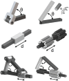
Fig. 1.
Fig. 1.
DNA origami mechanism design. Our approach to DNA origami mechanism design follows macroscopic machine design starting with isolated joints for angular (Top) or linear (Middle) motion. Joints can be integrated to achieve complex motion as shown here for a crank–slider mechanism (Bottom). Left shows macroscale solid models and Right shows their DNA origami counterparts. In the DNA origami designs, cylinders represent dsDNA helices.
Fig. 2.
Fig. 2.
DNA origami sliders. (A) The slider consists of two stiff components folded concentrically and connected only with ssDNA scaffold facilitating linear motion. Two versions of the slider were fabricated, one with short ssDNA connections (version 1) and one with long ssDNA connections (version 2). TEM images illustrate different conformations of version 2. Scale bar, 50 nm (B) Version 1 (shorter connections) is shown via TEM. Scale bar, 100 nm (C) A linear distribution of 14 nm was measured from 275 samples of version 1. (D) A wider linear distribution was measured from 251 samples of version 2. (E) The energy landscape was calculated for both versions from the linear distributions assuming Boltzmann energy weighting (scale bar indicates an energy scale of kBT). The lines show cubic spline fits to linear distributions. (F) The energy landscape was differentiated to determine the force required to hold each joint at any specific length.
Fig. 3.
Fig. 3.
DNA origami hinges. (A) The hinge consists of two stiff bundles of 18 dsDNA helices connected at one end by 6 ssDNA connections (white lines). Two versions of the hinge were fabricated. The short connections are 2 nt long in both designs and the long connections are 16 and 30 nt for hinge 1 and hinge 2, respectively. (B) TEM images of hinge 2 confirm well-folded structures and flexible motion in one angular degree of freedom. Scale bar, 100 nm. (C) The angular distribution of hinge 1, measured from 918 structures in TEM images, shows a torsionally stiff joint with an equilibrium angle of ∼85°. (D) The angular distribution of hinge 2, measured from 248 structures in TEM images, shows resistance to small (<40°) and large (>80°) angles with relative flexibility in the range between. (E) The energy landscape was calculated from the angular distributions assuming Boltzmann energy weighting (scale bar indicates an energy scale of kBT). The lines show cubic spline fits to angular distributions. (F) The energy landscape was differentiated to determine the torque required to hold each hinge at any specific angle.
Fig. 4.
Fig. 4.
DNA origami crank–slider coupling linear and rotational motion. (A) The mechanism incorporates three hinges and one slider joint using designs from Figs. 2 and 3 to achieve 2D motion. (B) TEM shows samples of the mechanisms. Scale bar, 100 nm. (C) The motion of the DNA origami crank–slider, illustrated by measurements of rotation vs. extension from TEM images for 56 samples (gray “x”s), follows the theoretical prediction (black line) for its rigid-body counterpart with some fluctuation about the ideally constrained motion path. TEM images on the right depict zoomed-in views of crank–sliders in different configurations along the motion path. Scale bar, 50 nm.
Fig. 5.
Fig. 5.
DNA origami mechanism with 3D motion. (A) The four-bar mechanism called a Bennett linkage traverses a complex 3D motion path between extreme configurations of an open frame (Top Left) or a compacted bundle (Bottom Left). (B) TEM images confirm well-folded structures. In the absence of “locking strands” the mechanism fluctuates freely along its motion path. Several structures in different conformations are highlighted. (C) A comparison of the motion quantified in terms of the projected internal angles demonstrates that the DNA origami mechanism closely follows the expected motion path for its rigid-body counterpart (black line). Conformations were measured for 52 structures (gray “x”s) (D) Structures were fixed in their fully expanded frame configuration and (E) in their fully compacted bundle configuration. Scale bar, 100 nm.
Fig. 6.
Fig. 6.
Actuation of DNA origami mechanisms. (A) Distributed actuation was designed with several connections along the length of the links to zipper the mechanism into a higher energy compacted configuration. The compacted mechanisms can be expanded after a second addition of ssDNA inputs via strand displacement. (B) TEM images show the freely fluctuating configuration before actuation with input strands. In the free configuration 9.9% of mechanisms appear in the bundle conformation. (C) DNA origami mechanisms were actuated by adding twofold excess of closing strands that connect overhangs on different arms. After actuating the forward process (closing), 93% of mechanisms are found in the compacted bundle configuration on TEM images. (D) The reverse process (expanding) is achieved by a second set of DNA inputs that removes the closing strands by DNA strand displacement. (E) Fluorescence quenching data (black) reveal the timescale of compacting to be t1/2,c = 55 s. (F) Expanding occurs on the timescale of t1/2,e = 49 s. Single- (blue) and double- (red) exponential fits are shown as dashed lines. Unconstrained, compacted, and expanded controls are shown in green. The expanded control exhibits lower fluorescence because structures are diluted by addition of actuation strands. Scale bar, 100 nm.

References

    1. Hamdi M, Ferreira A. Multiscale design and modeling of protein-based nanomechanisms for nanorobotics. Int J Robot Res. 2009;28(4):436–449.
    1. Mavroidis C, Dubey A, Yarmush ML. Molecular machines. Annu Rev Biomed Eng. 2004;6(1):363–395. - PubMed
    1. Ummat ADA, Sharma G, Mavroidis C. Bio-nanorobotics: State of the art and future challenges. In: Yarmush ML, editor. The Biomedical Engineering Handbook. CRC Press; Boca Raton, FL: 2006.
    1. Stephanopoulos N, Ortony JH, Stupp SI. Self-assembly for the synthesis of functional biomaterials. Acta Mater. 2013;61(3):912–930. - PMC - PubMed
    1. Seeman NC. Nanomaterials based on DNA. Annu Rev Biochem. 2010;79:65–87. - PMC - PubMed

Publication types