. 2015 Feb;4(2):146-55.
doi: 10.5966/sctm.2014-0181. Epub 2015 Jan 5.

Therapeutic Potential of Adipose-Derived SSEA-3-Positive Muse Cells for Treating Diabetic Skin Ulcers

Affiliations

Therapeutic Potential of Adipose-Derived SSEA-3-Positive Muse Cells for Treating Diabetic Skin Ulcers

Kahori Kinoshita et al. Stem Cells Transl Med. 2015 Feb.

Abstract

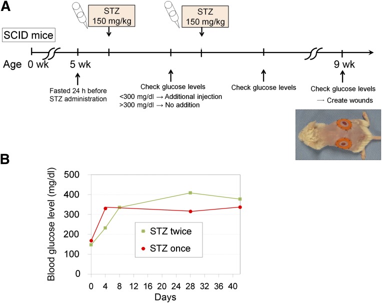
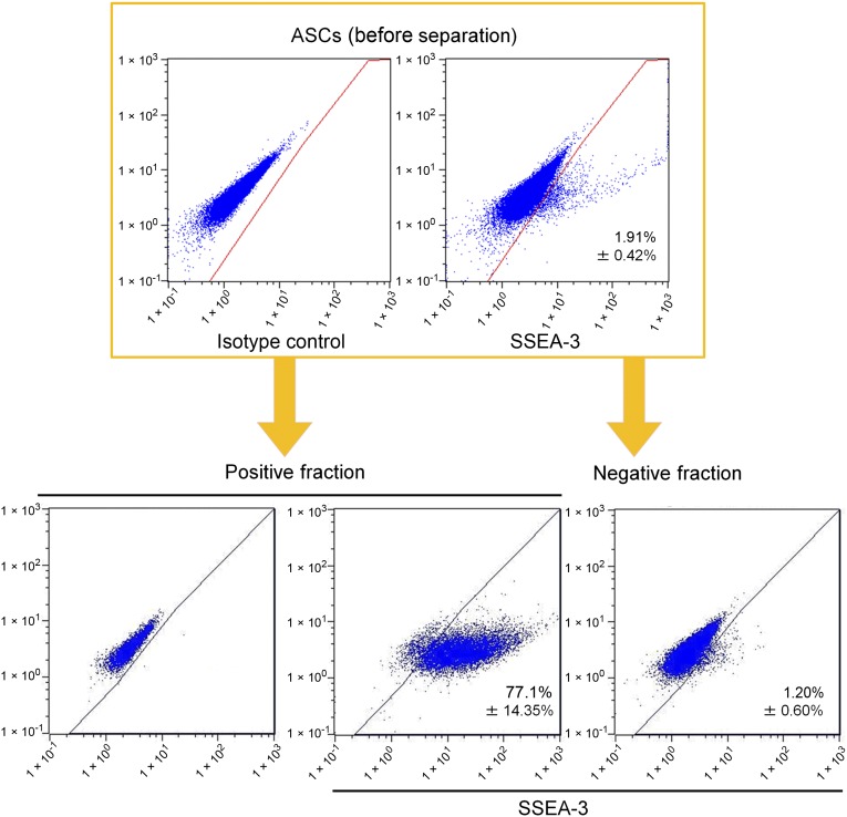
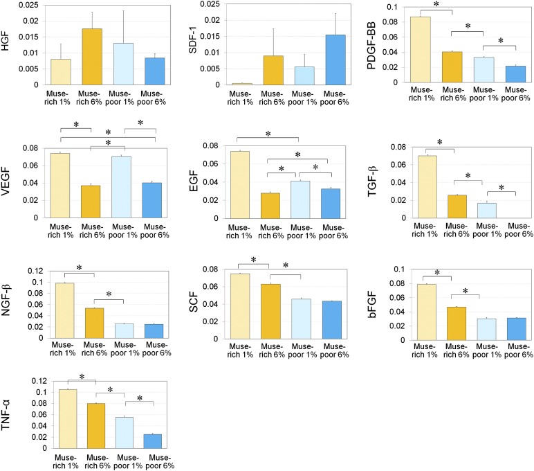
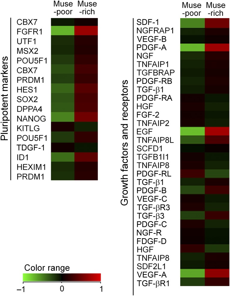
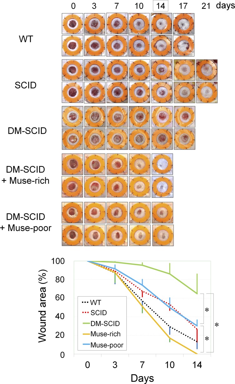
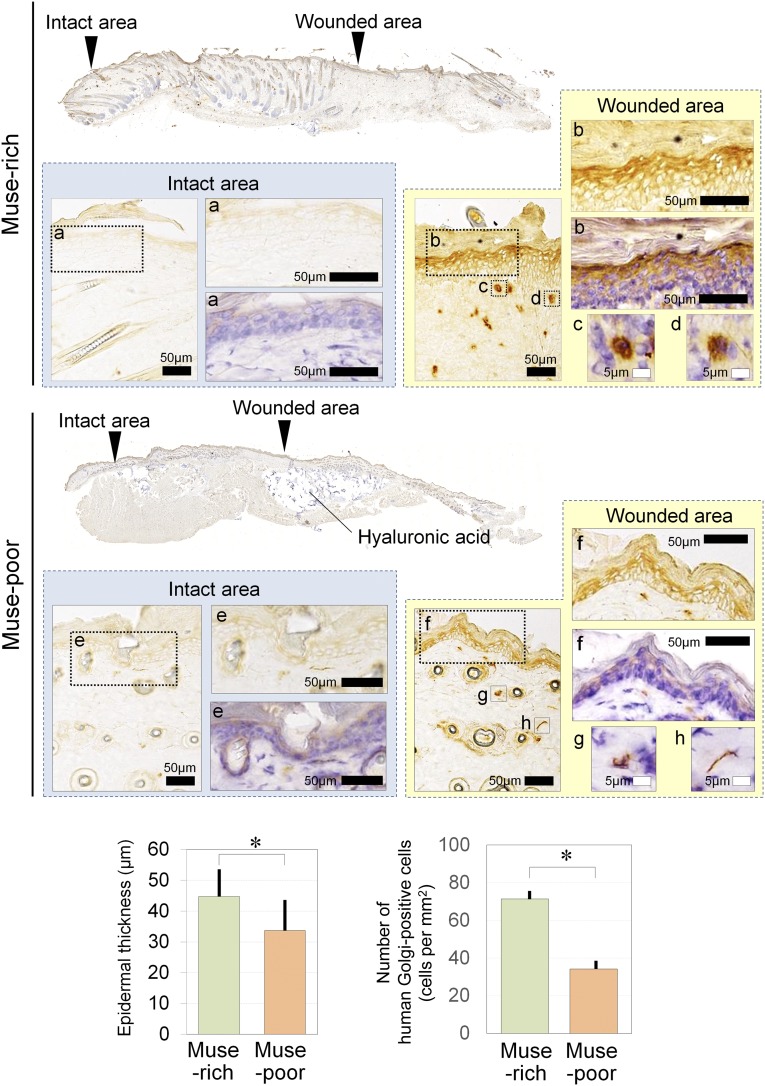
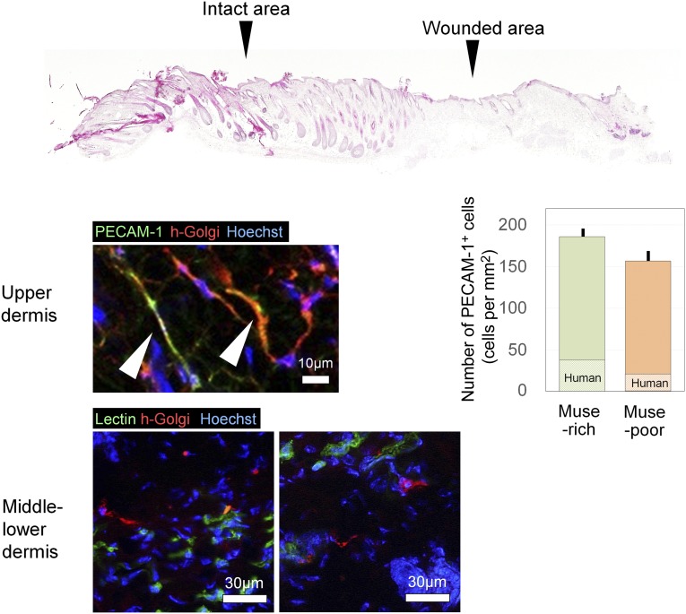
Stage-specific embryonic antigen-3 (SSEA-3)-positive multipotent mesenchymal cells (multilineage differentiating stress-enduring [Muse] cells) were isolated from cultured human adipose tissue-derived stem/stromal cells (hASCs) and characterized, and their therapeutic potential for treating diabetic skin ulcers was evaluated. Cultured hASCs were separated using magnetic-activated cell sorting into positive and negative fractions, a SSEA-3+ cell-enriched fraction (Muse-rich) and the remaining fraction (Muse-poor). Muse-rich hASCs showed upregulated and downregulated pluripotency and cell proliferation genes, respectively, compared with Muse-poor hASCs. These cells also released higher amounts of certain growth factors, particularly under hypoxic conditions, compared with Muse-poor cells. Skin ulcers were generated in severe combined immunodeficiency (SCID) mice with type 1 diabetes, which showed delayed wound healing compared with nondiabetic SCID mice. Treatment with Muse-rich cells significantly accelerated wound healing compared with treatment with Muse-poor cells. Transplanted cells were integrated into the regenerated dermis as vascular endothelial cells and other cells. However, they were not detected in the surrounding intact regions. Thus, the selected population of ASCs has greater therapeutic effects to accelerate impaired wound healing associated with type 1 diabetes. These cells can be achieved in large amounts with minimal morbidity and could be a practical tool for a variety of stem cell-depleted or ischemic conditions of various organs and tissues.

Keywords: Adipose; Adult stem cells; Cell culture; Endothelial cell; Hyaluronan; Mesenchymal stem cells; Stem cell transplantation; Tissue regeneration.


Figures

Figure 1.
Figure 1.
Preparation of immunodeficient diabetic mice. (A): We prepared immunodeficient mice with diabetes mellitus (DM) for a wound healing experiment. To induce type 1 DM, STZ was injected intraperitoneally into 5-week-old male SCID mice that had been fasted for 24 hours. Three days after STZ (150 mg/kg) administration, hyperglycemia (blood glucose >300 mg/dl) was examined. When hyperglycemia was not observed, another STZ (150 mg/kg) injection was given. Skin defects were created on the back of DM-induced SCID mice at 9 weeks of age. (B): Typical changes in blood glucose level are shown. Hyperglycemia was achieved after one or two STZ injections at approximately 75%. Abbreviations: SCID, severe combined immunodeficiency; STZ, streptozotocin.
Figure 2.
Figure 2.
Flow cytometry analyses for SSEA-3 expression before and after enrichment of Muse cells using magnetic-activated cell sorting (MACS). An example of flow cytometry analysis performed to measure SSEA-3+ cells before and after MACS cell enrichment and separation is shown. Cultured human ASCs were processed using MACS separation to obtain SSEA-3+ cells. The positive and negative cell fractions after MACS separation were used as Muse-rich and Muse-poor cell populations, respectively, in subsequent experiments. Abbreviations: ASCs, adipose tissue-derived stem/stromal cells; SSEA-3, stage-specific embryonic antigen-3.
Figure 3.
Figure 3.
Enzyme-linked immunosorbent assay (ELISA) analyses for growth factor production under hypoxic and normoxic conditions. The relative growth factor production values were measured with ELISA in Muse-rich and Muse-poor cell fractions cultured under hypoxic (1% O2) or normoxic (6% O2) conditions for 48 hours. The measured growth factors included HGF, SDF-1, PDGF-BB, VEGF, EGF, TGF-β, NGF-β, SCF, bFGF, and TNF-α. The y-axis indicates absorbance at 450 nm. The samples were collected from three independent experiments, and three replicates were used in each measurement. Data are presented as the mean ± SD (n = 3). ∗, p < .05. Abbreviations: bFGF, basic fibroblast growth factor; EGF, epidermal growth factor; HGF, hepatocyte growth factor; Muse, multilineage-differentiating stress-enduring; NGF-β, nerve growth factor-β; PDGF-BB, platelet-derived growth factor-BB; SCF, stem cell factor; SDF-1, stromal cell-derived factor 1; TGF-β, transforming growth factor-β; TNF-α, tumor necrosis factor-α; VEGF, vascular endothelial growth factor.
Figure 4.
Figure 4.
Microarray analyses of Muse-rich and Muse-poor cell populations. Heat maps for pluripotent markers, growth factors, and receptors indicate that pluripotent markers, including NANOG and FGFR1, were upregulated in the Muse-rich population compared with the Muse-poor population. The Muse-rich population also showed higher gene expression of growth factors such as PDGF-A, EGF, and VEGF-A (supplemental online Figure 1). Abbreviations: EGF, epidermal growth factor; FGFR1, fibroblast growth factor receptor 1; HGF, hepatocyte growth factor; Muse, multilineage-differentiating stress-enduring; NGF, nerve growth factor; PDGF-A, platelet-derived growth factor-A; SDF-1, stromal cell-derived factor 1; TGF, transforming growth factor; VEGF, vascular endothelial growth factor.
Figure 5.
Figure 5.
Wound healing of skin ulcers treated with Muse-rich and Muse-poor cell populations. Wound healing of skin defects (6 mm in diameter) was sequentially evaluated for up to 14 days. The wound area was photographed, and the percentage of the wound area to the original wound size was calculated using digital imaging software. Although SCID mice originally showed slightly delayed wound healing compared with their WT counterparts, the wound healing in the STZ-induced DM-SCID mice was more impaired than that in the SCID mice. Wound closure was significantly faster in the DM-SCID mice treated with the Muse-rich cell population than in Muse-poor-treated DM-SCID mice, which showed significantly better wound healing than the nontreated DM-SCID mice. Six mice were used in each group. ∗, p < .05. Abbreviations: DM-SCID, diabetes mellitus-induced SCID mice; Muse, multilineage-differentiating stress-enduring; SCID, severe combined immunodeficiency; STZ, streptozotocin; WT, wild-type.
Figure 6.
Figure 6.
Immunohistologic findings for human Golgi complex of diabetes mellitus-induced severe combined immunodeficiency wounds treated with Muse-rich or Muse-poor cell populations. Shown are high-magnification views of the intact epidermis (a), repaired epidermis (b), and wounded area (c, d) in Muse-rich sample. Also shown are high-magnification views of the intact epidermis (e), repaired epidermis area (f), and wounded area (g, h) in Muse-poor sample. Human Golgi complex-positive cells, which are equivalent to transplanted human cells, were observed in the epidermis and dermis of the wounded area in both Muse-rich and Muse-poor samples after 14 days. However, human Golgi complex-positive cells were not detected in the surrounding intact area in either group. Transplanted human cells were significantly more frequently detected in Muse-rich samples compared with Muse-poor samples (p = .0006). A significantly thicker epidermis was also noted in Muse-rich samples (p = .0053). ∗, p < .05. Abbreviation: Muse, multilineage-differentiating stress-enduring.
Figure 7.
Figure 7.
Immunohistologic findings of differentiation markers expressed by transplanted Muse-rich cells. Double immunohistochemistry was performed for human-specific proteins (human Golgi complex) and differentiation markers (PECAM-1 or isolectin) to characterize transplanted Muse-rich cells at day 14. Some cells expressing human Golgi complex were positive for PECAM-1 or isolectin, suggesting differentiation into vascular endothelial cells in the upper dermis; however, human Golgi complex-positive cells in the middle and lower dermis were negative for PECAM-1 and isolectin. The number of PECAM-1+ cells per microscopic field was counted, with no significant difference between the two groups (p = .144), although the ratio of human-derived cells was higher in the Muse-rich samples (p = .02). Abbreviations: h-Golgi, human Golgi complex; Muse, multilineage-differentiating stress-enduring; PECAM-1, platelet endothelial cell adhesion molecule-1.

Similar articles

Cited by

References

    1. Petersen BE, Bowen WC, Patrene KD, et al. Bone marrow as a potential source of hepatic oval cells. Science. 1999;284:1168–1170. - PubMed
    1. Harris RG, Herzog EL, Bruscia EM, et al. Lack of a fusion requirement for development of bone marrow-derived epithelia. Science. 2004;305:90–93. - PubMed
    1. Ren G, Chen X, Dong F, et al. Concise review: Mesenchymal stem cells and translational medicine: Emerging issues. Stem Cells Translational Medicine. 2012;1:51–58. - PMC - PubMed
    1. Amoh Y, Li L, Katsuoka K, et al. Multipotent nestin-positive, keratin-negative hair-follicle bulge stem cells can form neurons. Proc Natl Acad Sci USA. 2005;102:5530–5534. - PMC - PubMed
    1. Toma JG, Akhavan M, Fernandes KJ, et al. Isolation of multipotent adult stem cells from the dermis of mammalian skin. Nat Cell Biol. 2001;3:778–784. - PubMed

Publication types

MeSH terms

Substances