. 2015 Feb 20;290(8):4759-4771.
doi: 10.1074/jbc.M114.628412. Epub 2015 Jan 5.

Structure of neuroblastoma suppressor of tumorigenicity 1 (NBL1): insights for the functional variability across bone morphogenetic protein (BMP) antagonists

Affiliations

Structure of neuroblastoma suppressor of tumorigenicity 1 (NBL1): insights for the functional variability across bone morphogenetic protein (BMP) antagonists

Kristof Nolan et al. J Biol Chem. .

Abstract

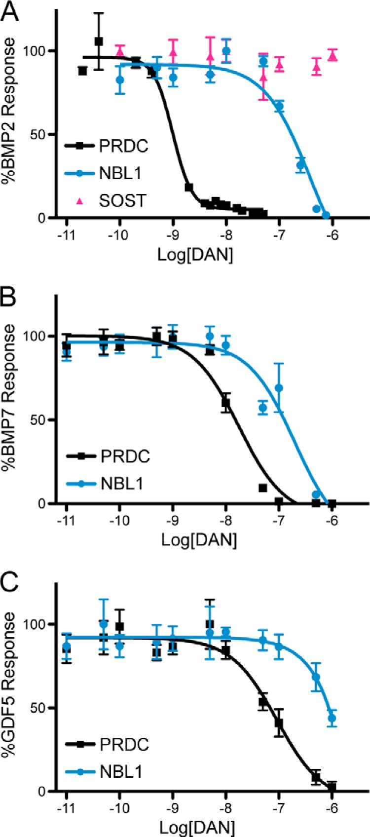
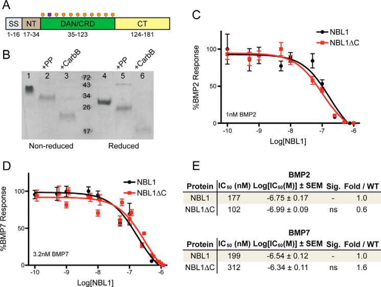
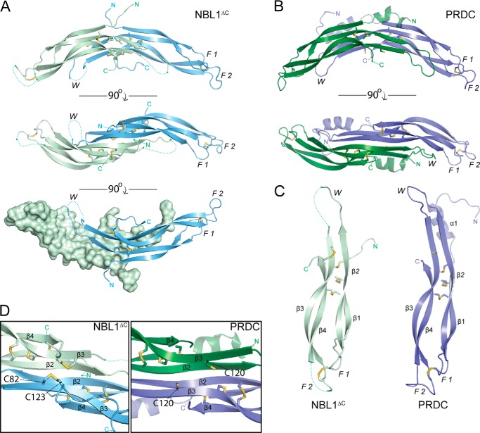
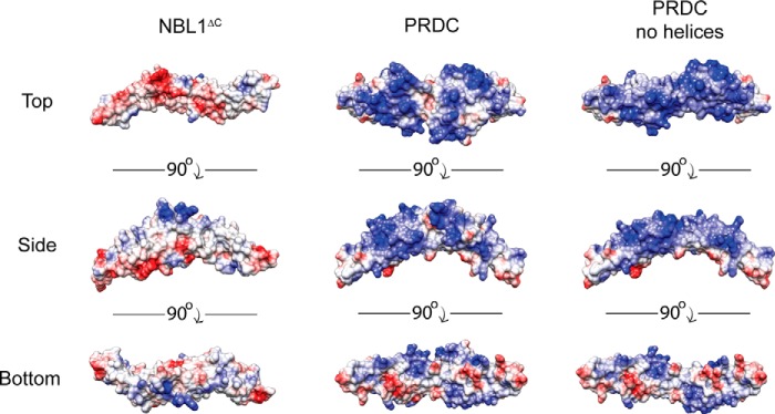
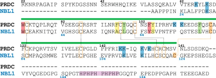
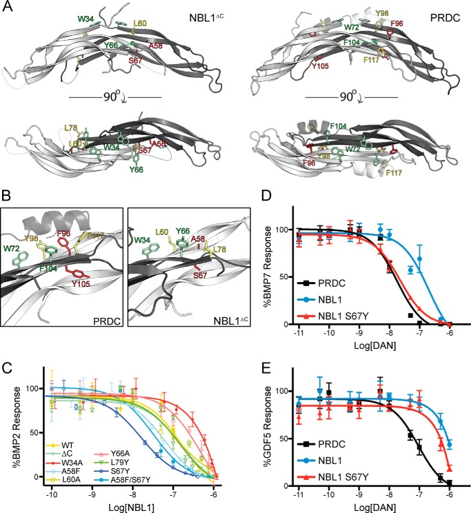
Bone morphogenetic proteins (BMPs) are antagonized through the action of numerous extracellular protein antagonists, including members from the differential screening-selected gene aberrative in neuroblastoma (DAN) family. In vivo, misregulation of the balance between BMP signaling and DAN inhibition can lead to numerous disease states, including cancer, kidney nephropathy, and pulmonary arterial hypertension. Despite this importance, very little information is available describing how DAN family proteins effectively inhibit BMP ligands. Furthermore, our understanding for how differences in individual DAN family members arise, including affinity and specificity, remains underdeveloped. Here, we present the structure of the founding member of the DAN family, neuroblastoma suppressor of tumorigenicity 1 (NBL1). Comparing NBL1 to the structure of protein related to Dan and Cerberus (PRDC), a more potent BMP antagonist within the DAN family, a number of differences were identified. Through a mutagenesis-based approach, we were able to correlate the BMP binding epitope in NBL1 with that in PRDC, where introduction of specific PRDC amino acids in NBL1 (A58F and S67Y) correlated with a gain-of-function inhibition toward BMP2 and BMP7, but not GDF5. Although NBL1(S67Y) was able to antagonize BMP7 as effectively as PRDC, NBL1(S67Y) was still 32-fold weaker than PRDC against BMP2. Taken together, this data suggests that alterations in the BMP binding epitope can partially account for differences in the potency of BMP inhibition within the DAN family.

Keywords: Bone Morphogenetic Protein (BMP); Differential Screening-aberrative in Neuroblastoma (DAN); Extracellular Antagonism; Mutagenesis; Neuroblastoma Suppressor of Tumorigenicity 1 (NBL1); Protein Related to Dan and Cerberus (PRDC); SOST; Structural Biology; Transforming Growth Factor β(TGF-B); X-ray Crystallography.


Figures

FIGURE 1.
FIGURE 1.
Comparison of BMP specificity and inhibition for PRDC and NBL1. PRDC and NBL1 (SOST only tested in A) were titrated against 1 nm BMP2 (A), 3.2 nm BMP7 (B), and 3.7 nm GDF5 (C) using a BMP responsive osteoblast cell line (BRITER) to measure inhibition and determine relative IC50 values. Experiments were performed in triplicate and the curves represent the average of three individual experiments (error bars represent ±S.E.).
FIGURE 2.
FIGURE 2.
Comparison of NBL1 and NBL1ΔC. A, schematic of the NBL1 protein. The protein can be separated into 4 regions: the signal sequence (SS), N terminus (NT), cysteine-rich or DAN domain (DAN/CRD), and C terminus (CT). Numbers under the diagram represent the amino acids in NBL1 composing each region. Orange circles represent cysteines and the purple box represents glycosylation. B, SDS-PAGE gel of purified NBL1 with the Myc-His6 tag used for purification in lanes 1 and 4, full-length NBL1 after remove of the purification tag using Prescission Protease (PP) in lanes 2 and 5, and NBL1 after cleavage using carboxypeptidase B (NBL1ΔC) in lanes 3 and 6. Proteins in lanes 4–6 were reduced using 10 mm β-mercaptoethanol. C and D, luciferase reporter assay testing the activity of NBL1 and NBL1ΔC against (C) 1 nm BMP2 and (D) 3.2 nm BMP7. E, table summarizing inhibition data for NBL1 and NBL1ΔC. IC50 values and significance were determined from Luciferase reporter assays using the Prism software package. NS, not significant based upon a p value of 0.05.
FIGURE 3.
FIGURE 3.
Crystal structure of NBL1ΔC. A, ribbon representation of the NBL1ΔC dimer with the Chain A monomer shown in pale green and Chain B monomer shown in pale blue. Sticks represent disulfide bonds with sulfurs colored yellow-orange. Dotted lines represent areas that cannot be resolved in the electron density and connect amino acids according to the primary protein sequence. Different views (from top to bottom) show the dimer rotated 90° about the horizontal axis. F1, finger 1; F2, finger 2; W, wrist region. B, ribbon representation of the PRDC dimer crystal structure (Protein Data Bank code 4JPH, Ref. 16). C, comparison of the NBL1ΔC and PRDC monomer structures. β-Strands are labeled in each monomer (β1-β4) in order from the N terminus. D, view of the bottom, concave surfaces of NBL1ΔC (left) and PRDC (right), highlighting their cystine knots. As can be seen, the fifth disulfide bond in NBL1ΔC links the final cysteine of the protein to the synonymous free cysteine in PRDC and reiterates that these proteins do not form covalently attached dimers.
FIGURE 4.
FIGURE 4.
Comparison of NBL1ΔC and PRDC electrostatics. Three views of NBL1ΔC, PRDC, and PRDC lacking its N-terminal helices, depicting the electrostatic surface potential of these proteins from the top, bottom, and side perspectives. Surface potential was calculated using APBS and the proteins are colored based on a scale from −10 to 10 kbT/ec (red to blue).
FIGURE 5.
FIGURE 5.
Sequence alignment of NBL1 and PRDC. Alignment of the mouse PRDC (top) and human NBL1 (bottom) primary sequences (excluding signaling sequences). Numbers represent the amino acid for the corresponding antagonist (black for PRDC and blue for NBL1). Green bar over the alignment indicates the location of the DAN domain. Black dotted lines show disulfide bonds shared between both proteins. Orange dotted line represents the unique fifth disulfide bond present in NBL1. Lysines highlighted in blue are suggested to be important for heparin binding. Histidine-proline repeat in the C terminus of NBL1, believed to be important for metal binding, is highlighted in purple. Cysteines are highlighted orange. Amino acids highlighted in green, yellow, and red have been shown to be important for PRDC to bind to BMP2. Those highlighted in yellow are not conserved between PRDC and NBL1, whereas those in green and red are partially or well conserved, respectively.
FIGURE 6.
FIGURE 6.
Mutagenesis studies on NBL1. Ribbon representation of NBL1ΔC (A) and PRDC (B) showing amino acids (stick representation) selected for our mutagenesis studies based upon data obtained for characterizing the PRDC BMP-binding epitope. Each of these amino acids in a previous study was found to be important for mediating BMP inhibition in PRDC. Residues selected for mutagenesis that are poorly conserved between NBL1 and PRDC and colored in red, whereas those that are moderately and completely conserved are depicted in yellow and green, respectively. B, close up view of the residues selected for these studies and depicting the known portion of the PRDC BMP-binding epitope. C, luciferase reporter assay showing titration of various NBL1 mutants against 1 nm BMP2. The wild-type NBL1 protein and mutants with similar IC50 values are colored green to yellow, whereas those that resulted in reduction of inhibition compared with wild-type are colored red and those that result in improved inhibition are colored blue. D and E, luciferase reporter assay results comparing the activities of PRDC, NBL1 and NBL1S67Y when titrated against 3.2 nm BMP7 (D) and 3.7 nm GDF5 (E).
FIGURE 7.
FIGURE 7.
Analysis of NBL1 and NBL1 mutants in vivo. In vivo BMP inhibition activity of NBL1 and various mutants tested during Xenopus embryo development. The purified proteins (0.5, 2, and 10 μm) were injected into the blastocoel cavity of stage 9 embryos. A, bar graph represents embryos that were scored for defects in BMP-dependent axial development at stage 35 using the standard dorsoanterior index and classified into different subgroups based upon the severity of posterior truncation. Each bar represents 100% percent of the embryos tested where cyan represents the percent of the population with a normal phenotype, yellow represents a mild phenotype (mild tail truncation), and red represents a severe axial truncation. Images show representative examples of each described phenotype. B, embryos at stage 20 (ventral views) were evaluated by in situ hybridization for mRNA expression of the direct BMP-target gene sizzled. In control embryos sizzled expression is detected in the ventral mesoderm as a result of endogenous BMP signaling (BSA/PBS control), whereas strong inhibition of endogenous BMP signaling results in very little to no sizzled expression (DMH-1 control). Each protein was tested in at least 3 separate injection experiments, with 80–100 embryos tested in total for each.

References

    1. Bragdon B., Moseychuk O., Saldanha S., King D., Julian J., Nohe A. (2011) Bone morphogenetic proteins: a critical review. Cell Signal. 23, 609–620 - PubMed
    1. Cai J., Pardali E., Sánchez-Duffhues G., ten Dijke P. (2012) BMP signaling in vascular diseases. FEBS Lett. 586, 1993–2002 - PubMed
    1. Walsh D. W., Godson C., Brazil D. P., Martin F. (2010) Extracellular BMP-antagonist regulation in development and disease: tied up in knots. Trends Cell Biol. 20, 244–256 - PubMed
    1. Rider C. C., Mulloy B. (2010) Bone morphogenetic protein and growth differentiation factor cytokine families and their protein antagonists. Biochem. J. 429, 1–12 - PubMed
    1. Tardif G., Pelletier J.-P., Boileau C., Martel-Pelletier J. (2009) The BMP antagonists Follistatin and Gremlin in normal and early osteoarthritic cartilage: an immunohistochemical study. Osteoarthr. Cartil. 17, 263–270 - PubMed

Publication types

MeSH terms

Associated data

LinkOut - more resources