Review
. 2015 Jan;8(1):1-16.
doi: 10.1242/dmm.017400.

How informative is the mouse for human gut microbiota research?

Affiliations
Review

How informative is the mouse for human gut microbiota research?

Thi Loan Anh Nguyen et al. Dis Model Mech. 2015 Jan.

Abstract

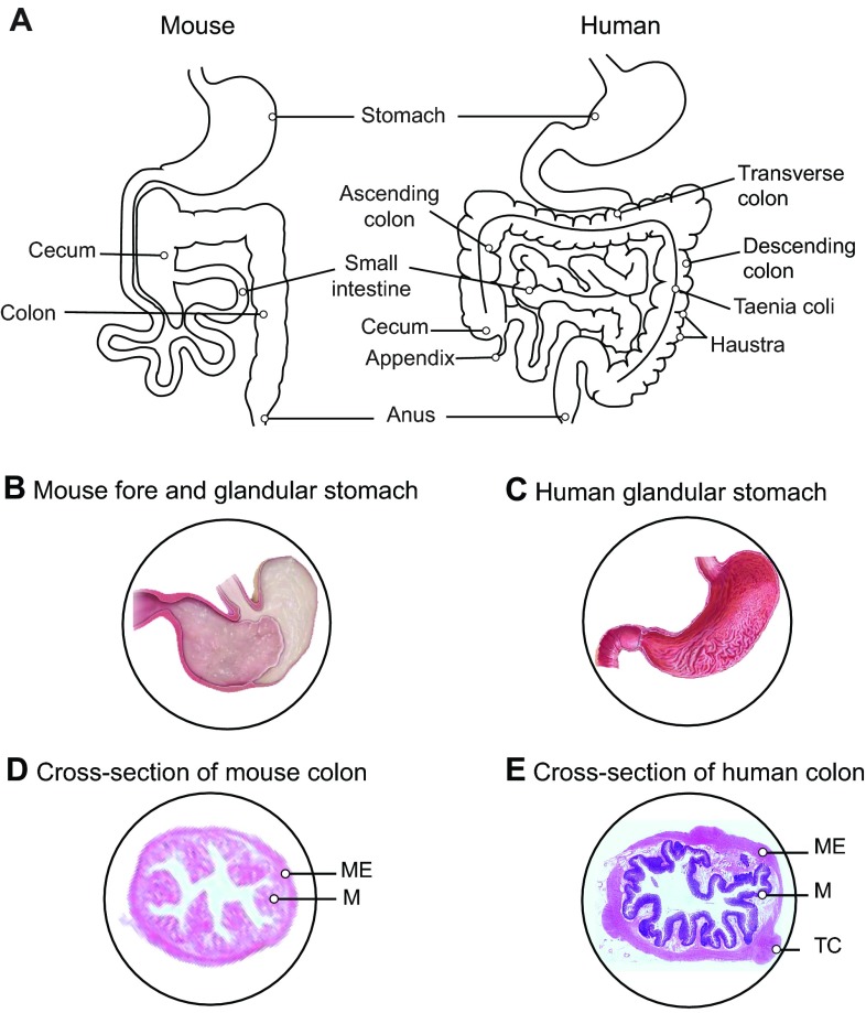
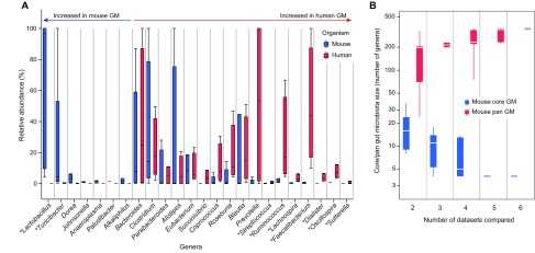
The microbiota of the human gut is gaining broad attention owing to its association with a wide range of diseases, ranging from metabolic disorders (e.g. obesity and type 2 diabetes) to autoimmune diseases (such as inflammatory bowel disease and type 1 diabetes), cancer and even neurodevelopmental disorders (e.g. autism). Having been increasingly used in biomedical research, mice have become the model of choice for most studies in this emerging field. Mouse models allow perturbations in gut microbiota to be studied in a controlled experimental setup, and thus help in assessing causality of the complex host-microbiota interactions and in developing mechanistic hypotheses. However, pitfalls should be considered when translating gut microbiome research results from mouse models to humans. In this Special Article, we discuss the intrinsic similarities and differences that exist between the two systems, and compare the human and murine core gut microbiota based on a meta-analysis of currently available datasets. Finally, we discuss the external factors that influence the capability of mouse models to recapitulate the gut microbiota shifts associated with human diseases, and investigate which alternative model systems exist for gut microbiota research.

Keywords: Gut microbiota; Humanized mouse models; Mouse core gut microbiota; Mouse models; Mouse pan-gut microbiota.


Figures

Fig. 1.
Fig. 1.
Gross anatomy of the human and the mouse gastrointestinal tract. (A) The human colon is divided into different sections (i.e. ascending, transverse and descending colon) with the presence of taenia coli and compartmentalization in haustra, which are absent in the mouse colon. The human stomach is lined with a glandular mucosa (C) that secretes gastric acid, whereas the mouse stomach is divided in two regions – a glandular stomach and a non-glandular or fore-stomach (B). The mouse glandular stomach is responsible for secreting gastric acid, whereas the non-glandular stomach functions as a temporary site of food storage and digestion. (E) Cross-section of a human colon, which has a thicker muscular wall and mucosa compared with the mouse colon (D). M, mucosa; ME, muscularis externa; TC, taenia coli. Panels B and C are reproduced from “Comparative anatomy and histology: A mouse and human atlas” by Piper M. Treuting and Suzy Dintzis, 2012, with permission from Elsevier. D is reproduced from the website http://theses.ulaval.ca/archimede/fichiers/24866/ch07.html with the author’s permission. E is re-used from www.anatomyatlases.org with the author’s permission.
Fig. 2.
Fig. 2.
Anatomical structures of the intestinal wall in mice and in humans. A and B are taken at the same magnification (20×) and show a section of the small intestinal wall in mice (A) and humans (B), illustrating that mouse intestinal villi (A) are taller than those in humans (B). C, intestinal crypts; G, goblet cells; L, lamina propria; MM, muscularis mucosae; P, Paneth cells; SM, submucosa; V, villi. The images are reproduced from “Comparative anatomy and histology: A mouse and human atlas” by Piper M. Treuting and Suzy Dintzis, 2012, with permission from Elsevier.
Fig. 3.
Fig. 3.
Meta-analysis of mouse and human fecal microbiota based on published 16S rDNA sequencing data. (A) Comparison of human and mouse healthy adult gut microbiota. The relative abundances of genera in the gut microbiota of both organisms [four human datasets (Human Microbiome Project Consortium, 2012; Yatsunenko et al., 2012) and five mouse datasets (Nagy-Szakal et al., 2012; Riboulet-Bisson et al., 2012; Ubeda et al., 2013; Ward et al., 2012; Zenewicz et al., 2013)] are ordered according to their overrepresentation in either mouse or human gut microbiota (non-parametric Wilcoxon Z score). Genera with significant differences (P<0.05) between human and mouse gut microbiota are annotated with an asterisk. Note that none of these differences are significant after multiple testing corrections. Note that only five mouse datasets are used in this comparison because the dataset from Cho et al. (Cho et al., 2012) does not include data for abundance and thus cannot be used for comparison of relative abundances between mouse and human gut microbiota. (B) Mouse core and pan-gut microbiota size in all possible combinations of the six mouse gut microbiota datasets (Cho et al., 2012; Nagy-Szakal et al., 2012; Riboulet-Bisson et al., 2012; Ubeda et al., 2013; Ward et al., 2012; Zenewicz et al., 2013). The pan-gut microbiota is the set of genera found at least once in any of the datasets compared (union set), whereas the core gut microbiota is the set of genera found in all compared datasets (intersection set). It should be noted that Zenewicz’s dataset overlaps poorly with the others.
Fig. 4.
Fig. 4.
Histological features of murine DSS-induced colitis. (A,B) Histology images of colon cross-sections from control (A) and dextran sulfate sodium (DSS)-treated (B) mice. The diagram under each panel illustrates an outline of a mouse colon cross-section with boxes A and B indicating the histological regions shown in panels A and B, respectively. Mir29-knockout mice from a Black6 background were treated either with water (control animals) or with 1.5% DSS for 8 days prior to examination. Whereas the cross-section of the colon wall from a control mouse shows normal morphology (A), the colon of the DSS-treated mouse (B) shows alterations in the mucosal epithelium such as cell necrosis, and loss of structure of both villi and intestinal crypts (C; indicated by arrowhead). In addition, infiltration of immune cells such as macrophages, neutrophils, eosinophils and lymphocytes (indicated with arrowheads) is found in the colonic lamina propria (L) and submucosa (SM), indicative of the inflammatory status of the DSS-treated mouse. C, intestinal crypts; G, goblet cells; L, lamina propria; ME, muscularis externa; MM, muscularis mucosae; MT: muscularis tunics; SM: submucosa; V: villi.

References

    1. Akaza H. (2012). Prostate cancer chemoprevention by soy isoflavones: role of intestinal bacteria as the “second human genome”. Cancer Sci. 103, 969–975. - PMC - PubMed
    1. Alpert C., Sczesny S., Gruhl B., Blaut M. (2008). Long-term stability of the human gut microbiota in two different rat strains. Curr. Issues Mol. Biol. 10, 17–24. - PubMed
    1. Andoh A., Kuzuoka H., Tsujikawa T., Nakamura S., Hirai F., Suzuki Y., Matsui T., Fujiyama Y., Matsumoto T. (2012). Multicenter analysis of fecal microbiota profiles in Japanese patients with Crohn’s disease. J. Gastroenterol. 47, 1298–1307. - PubMed
    1. Arumugam M., Raes J., Pelletier E., Le Paslier D., Yamada T., Mende D. R., Fernandes G. R., Tap J., Bruls T., Batto J.-M., et al. MetaHIT Consortium (2011). Enterotypes of the human gut microbiome. Nature 473, 174–180. - PMC - PubMed
    1. Bäckhed F., Manchester J. K., Semenkovich C. F., Gordon J. I. (2007). Mechanisms underlying the resistance to diet-induced obesity in germ-free mice. Proc. Natl. Acad. Sci. USA 104, 979–984. - PMC - PubMed