A head-mounted display-based personal integrated-image monitoring system for transurethral resection of the prostate
- PMID: 25562008
- PMCID: PMC4280403
- DOI: 10.5114/wiitm.2014.43040
A head-mounted display-based personal integrated-image monitoring system for transurethral resection of the prostate
Abstract
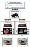
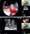
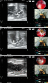
The head-mounted display (HMD) is a new image monitoring system. We developed the Personal Integrated-image Monitoring System (PIM System) using the HMD (HMZ-T2, Sony Corporation, Tokyo, Japan) in combination with video splitters and multiplexers as a surgical guide system for transurethral resection of the prostate (TURP). The imaging information obtained from the cystoscope, the transurethral ultrasonography (TRUS), the video camera attached to the HMD, and the patient's vital signs monitor were split and integrated by the PIM System and a composite image was displayed by the HMD using a four-split screen technique. Wearing the HMD, the lead surgeon and the assistant could simultaneously and continuously monitor the same information displayed by the HMD in an ergonomically efficient posture. Each participant could independently rearrange the images comprising the composite image depending on the engaging step. Two benign prostatic hyperplasia (BPH) patients underwent TURP performed by surgeons guided with this system. In both cases, the TURP procedure was successfully performed, and their postoperative clinical courses had no remarkable unfavorable events. During the procedure, none of the participants experienced any HMD-wear related adverse effects or reported any discomfort.
Keywords: cystoscopes; data display; prostatic hyperplasia; transurethral resection of prostate.
Figures


Similar articles
-
Integrated image monitoring system using head-mounted display for gasless single-port clampless partial nephrectomy.Wideochir Inne Tech Maloinwazyjne. 2014 Dec;9(4):634-7. doi: 10.5114/wiitm.2014.44155. Epub 2014 Jul 22. Wideochir Inne Tech Maloinwazyjne. 2014. PMID: 25562006 Free PMC article.
-
Head-mounted display for a personal integrated image monitoring system: ureteral stent placement.Urol Int. 2015;94(1):117-20. doi: 10.1159/000356987. Epub 2014 Aug 13. Urol Int. 2015. PMID: 25139526
-
Patient's Self-monitoring of Transurethral Surgical Images Using a Head-mounted Display.Urol Case Rep. 2015 Jan 6;3(2):27-9. doi: 10.1016/j.eucr.2014.12.005. eCollection 2015 Mar. Urol Case Rep. 2015. PMID: 26793491 Free PMC article.
-
Economic Value of the Transurethral Resection in Saline System for Treatment of Benign Prostatic Hyperplasia in England and Wales: Systematic Review, Meta-analysis, and Cost-Consequence Model.Eur Urol Focus. 2018 Mar;4(2):270-279. doi: 10.1016/j.euf.2016.03.002. Epub 2016 Mar 23. Eur Urol Focus. 2018. PMID: 28753756
-
Randomised evaluation of alternative electrosurgical modalities to treat bladder outflow obstruction in men with benign prostatic hyperplasia.Health Technol Assess. 2005 Feb;9(4):iii-iv, 1-30. doi: 10.3310/hta9040. Health Technol Assess. 2005. PMID: 15698525 Review.
Cited by
-
A review of wearable technology in medicine.J R Soc Med. 2016 Oct;109(10):372-380. doi: 10.1177/0141076816663560. J R Soc Med. 2016. PMID: 27729595 Free PMC article. Review.
-
Integrated image navigation system using head-mounted display in "RoboSurgeon" endoscopic radical prostatectomy.Wideochir Inne Tech Maloinwazyjne. 2014 Dec;9(4):613-8. doi: 10.5114/wiitm.2014.44135. Epub 2014 Jul 19. Wideochir Inne Tech Maloinwazyjne. 2014. PMID: 25562001 Free PMC article.
-
A three-dimensional head-mounted display system (RoboSurgeon system) for gasless laparoendoscopic single-port partial cystectomy.Wideochir Inne Tech Maloinwazyjne. 2014 Dec;9(4):638-43. doi: 10.5114/wiitm.2014.44407. Epub 2014 Aug 3. Wideochir Inne Tech Maloinwazyjne. 2014. PMID: 25562007 Free PMC article.
-
The Use of Head-Worn Displays for Vital Sign Monitoring in Critical and Acute Care: Systematic Review.JMIR Mhealth Uhealth. 2021 May 11;9(5):e27165. doi: 10.2196/27165. JMIR Mhealth Uhealth. 2021. PMID: 33973863 Free PMC article.
-
Integrated image monitoring system using head-mounted display for gasless single-port clampless partial nephrectomy.Wideochir Inne Tech Maloinwazyjne. 2014 Dec;9(4):634-7. doi: 10.5114/wiitm.2014.44155. Epub 2014 Jul 22. Wideochir Inne Tech Maloinwazyjne. 2014. PMID: 25562006 Free PMC article.
References
-
- Oelke M, Bachmann A, Descazeaud A, et al. EAU guidelines on the treatment and follow-up of non-neurogenic male lower urinary tract symptoms including benign prostatic obstruction. Eur Urol. 2013;64:118–40. - PubMed
-
- Aliaev Iu G, Grigorian VA, Tsarichenko DG, et al. Transurethral electroresection of prostatic adenoma under transrectal ultrasonic control. Urologiia (Moscow, Russia: 1999) 2006;3:8–12. - PubMed
-
- Liu D, Jenkins SA, Sanderson PM, et al. Monitoring with head-mounted displays in general anesthesia: a clinical evaluation in the operating room. Anesth Analg. 2010;110:1032–8. - PubMed
Publication types
LinkOut - more resources
Full Text Sources
Other Literature Sources
