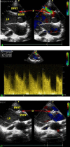
Minimally invasive transthoracic device closure of an acquired sinus of valsalva-right ventricle fistula in a pediatric patient
- PMID: 25562029
- PMCID: PMC4276590
Minimally invasive transthoracic device closure of an acquired sinus of valsalva-right ventricle fistula in a pediatric patient
Abstract
Background: Sinus of Valsalva-right ventricle fistula is a recognized but very rare complication after surgical repair of subaortic ventricular septal defect. Surgical repair with cardiopulmonary bypass and percutaneous transcatheter closure guided by x-ray has been the traditional treatment for fistula of sinus of Valsalva.
Case presentation: Recently, we have used a novel approach, that avoids the need for either secondary open surgical repair or radiation exposure; that is, minimally invasive transthoracic device closure guided by transesophageal echocardiography to occlude an acquired sinus of Valsalva-right ventricle fistula in a 4-year-old patient.
Conclusion: To our knowledge, there have been no prior cases reported of this technique applied to close an acquired sinus of Valsalva-right ventricle fistula. This report aims to provide a detailed description of the procedure.
Keywords: Fistula of Sinus of Valsalva; Minimally invasive; Device Closure; Transesophageal Echocardiography.
Figures


References
-
- Matsushita T, Masuda S, Inoue T, et al. Perforation of sinus Valsalva 10 years after repair of ventricular septal defect. Asian Cardiovasc Thorac Ann. 2012;20(3):353. - PubMed
-
- Sarikaya S, Adademir T, Elibol A, et al. Surgery for ruptured sinus of Valsalva aneurysm: 25-year experience with 55 patients. Eur J Cardiothorac Surg. 2013;43(3):591–6. - PubMed
-
- Khoury A, Khatib I, Halabi M, et al. Transcatheter closure of ruptured sinus of Valsalva aneurysm. Catheter Cardiovasc Interv. 2010;76(5):774–6. - PubMed
Publication types
LinkOut - more resources
Full Text Sources
