

Intrinsically disordered C-terminal tails of E. coli single-stranded DNA binding protein regulate cooperative binding to single-stranded DNA
- PMID: 25562210
- PMCID: PMC4419694
- DOI: 10.1016/j.jmb.2014.12.020
Intrinsically disordered C-terminal tails of E. coli single-stranded DNA binding protein regulate cooperative binding to single-stranded DNA
Abstract
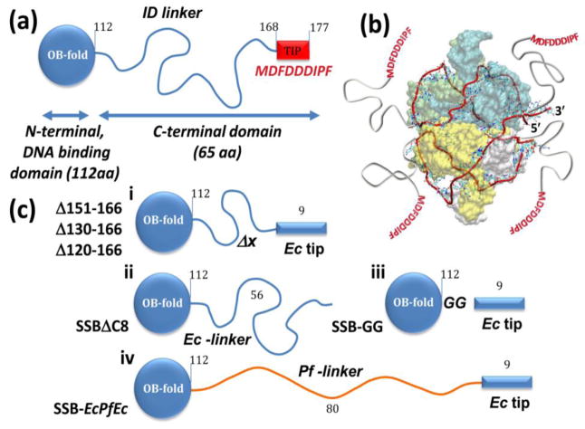
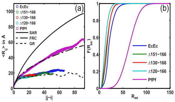
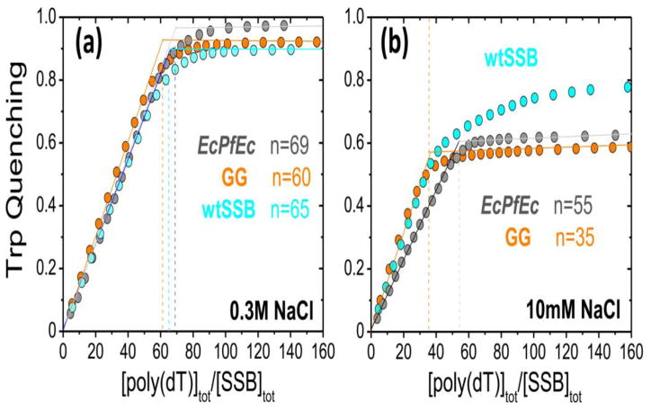
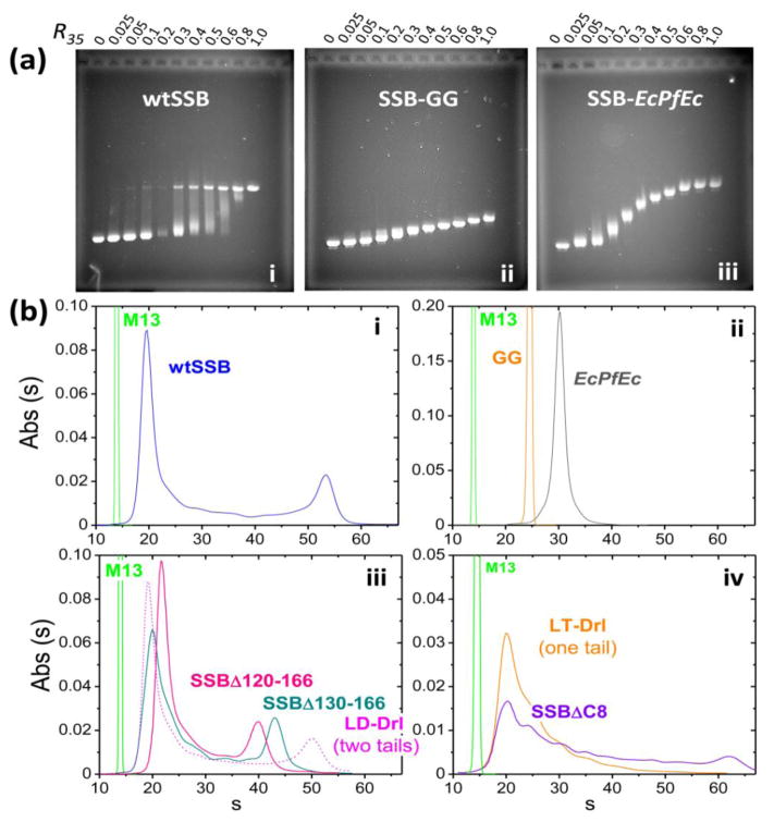
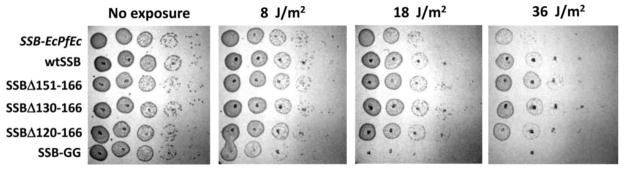
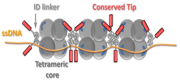
The homotetrameric Escherichia coli single-stranded DNA binding protein (SSB) plays a central role in DNA replication, repair and recombination. E. coli SSB can bind to long single-stranded DNA (ssDNA) in multiple binding modes using all four subunits [(SSB)65 mode] or only two subunits [(SSB)35 binding mode], with the binding mode preference regulated by salt concentration and SSB binding density. These binding modes display very different ssDNA binding properties with the (SSB)35 mode displaying highly cooperative binding to ssDNA. SSB tetramers also bind an array of partner proteins, recruiting them to their sites of action. This is achieved through interactions with the last 9 amino acids (acidic tip) of the intrinsically disordered linkers (IDLs) within the four C-terminal tails connected to the ssDNA binding domains. Here, we show that the amino acid composition and length of the IDL affects the ssDNA binding mode preferences of SSB protein. Surprisingly, the number of IDLs and the lengths of individual IDLs together with the acidic tip contribute to highly cooperative binding in the (SSB)35 binding mode. Hydrodynamic studies and atomistic simulations suggest that the E. coli SSB IDLs show a preference for forming an ensemble of globular conformations, whereas the IDL from Plasmodium falciparum SSB forms an ensemble of more extended random coils. The more globular conformations correlate with cooperative binding.
Keywords: DNA repair; DNA replication; cooperativity; regulation; simulations.
Copyright © 2015 Elsevier Ltd. All rights reserved.
Figures
Similar articles
-
Molecular insights into the prototypical single-stranded DNA-binding protein from E. coli.Crit Rev Biochem Mol Biol. 2024 Feb-Apr;59(1-2):99-127. doi: 10.1080/10409238.2024.2330372. Epub 2024 May 21. Crit Rev Biochem Mol Biol. 2024. PMID: 38770626 Free PMC article. Review.
-
Regulation of single-stranded DNA binding by the C termini of Escherichia coli single-stranded DNA-binding (SSB) protein.J Biol Chem. 2010 May 28;285(22):17246-52. doi: 10.1074/jbc.M110.118273. Epub 2010 Apr 1. J Biol Chem. 2010. PMID: 20360609 Free PMC article.
-
The intrinsically disordered linker in the single-stranded DNA-binding protein influences DNA replication restart and recombination pathways in Escherichia coli K-12.J Bacteriol. 2024 Apr 18;206(4):e0033023. doi: 10.1128/jb.00330-23. Epub 2024 Mar 12. J Bacteriol. 2024. PMID: 38470036 Free PMC article.
-
Regulation of Nearest-Neighbor Cooperative Binding of E. coli SSB Protein to DNA.Biophys J. 2019 Dec 3;117(11):2120-2140. doi: 10.1016/j.bpj.2019.09.047. Epub 2019 Oct 28. Biophys J. 2019. PMID: 31708161 Free PMC article.
-
Escherichia coli single-stranded DNA-binding protein: multiple DNA-binding modes and cooperativities.Annu Rev Biochem. 1994;63:527-70. doi: 10.1146/annurev.bi.63.070194.002523. Annu Rev Biochem. 1994. PMID: 7979247 Review.
Cited by
-
Glutamate promotes SSB protein-protein Interactions via intrinsically disordered regions.J Mol Biol. 2017 Sep 1;429(18):2790-2801. doi: 10.1016/j.jmb.2017.07.021. Epub 2017 Aug 3. J Mol Biol. 2017. PMID: 28782560 Free PMC article.
-
Using microsecond single-molecule FRET to determine the assembly pathways of T4 ssDNA binding protein onto model DNA replication forks.Proc Natl Acad Sci U S A. 2017 May 2;114(18):E3612-E3621. doi: 10.1073/pnas.1619819114. Epub 2017 Apr 17. Proc Natl Acad Sci U S A. 2017. PMID: 28416680 Free PMC article.
-
Probing E. coli SSB protein-DNA topology by reversing DNA backbone polarity.Biophys J. 2021 Apr 20;120(8):1522-1533. doi: 10.1016/j.bpj.2021.02.025. Epub 2021 Feb 23. Biophys J. 2021. PMID: 33636169 Free PMC article.
-
DNA synthesis determines the binding mode of the human mitochondrial single-stranded DNA-binding protein.Nucleic Acids Res. 2017 Jul 7;45(12):7237-7248. doi: 10.1093/nar/gkx395. Nucleic Acids Res. 2017. PMID: 28486639 Free PMC article.
-
Molecular insights into the prototypical single-stranded DNA-binding protein from E. coli.Crit Rev Biochem Mol Biol. 2024 Feb-Apr;59(1-2):99-127. doi: 10.1080/10409238.2024.2330372. Epub 2024 May 21. Crit Rev Biochem Mol Biol. 2024. PMID: 38770626 Free PMC article. Review.
References
-
- Chase JW, Williams KR. Single-stranded DNA binding proteins required for DNA replication. Annu Rev Biochem. 1986;55:103–36. - PubMed
-
- Lohman TM, Ferrari ME. Escherichia coli single-stranded DNA-binding protein: multiple DNA-binding modes and cooperativities. Annu Rev Biochem. 1994;63:527–70. - PubMed
-
- Wold MS. Replication protein A: a heterotrimeric, single-stranded DNA-binding protein required for eukaryotic DNA metabolism. Annu Rev Biochem. 1997;66:61–92. - PubMed
Publication types
MeSH terms
Substances
Grants and funding
LinkOut - more resources
Full Text Sources
Other Literature Sources
Molecular Biology Databases
