. 2015 Feb;125(2):539-50.
doi: 10.1172/JCI76887. Epub 2014 Dec 22.

The HMGB1/RAGE axis triggers neutrophil-mediated injury amplification following necrosis

The HMGB1/RAGE axis triggers neutrophil-mediated injury amplification following necrosis

Peter Huebener et al. J Clin Invest. 2015 Feb.

Erratum in

Expression of concern in

Abstract

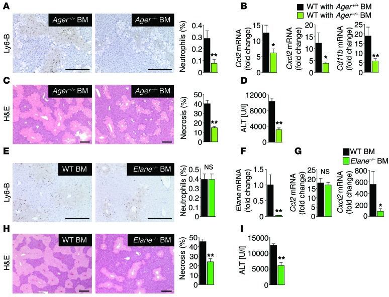
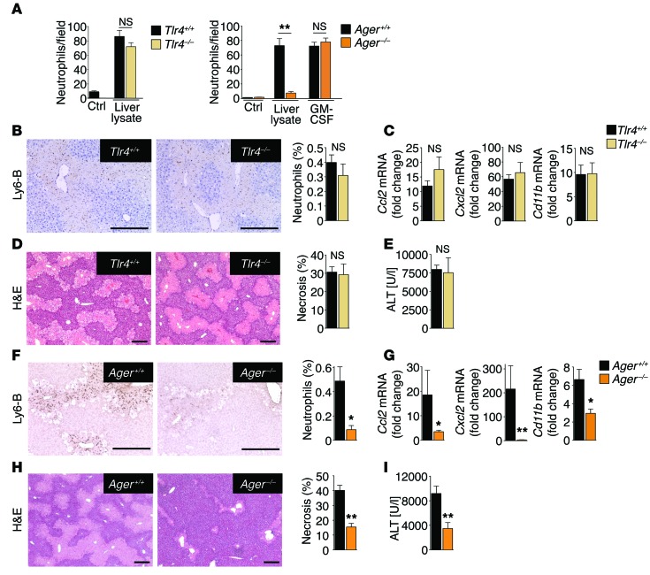
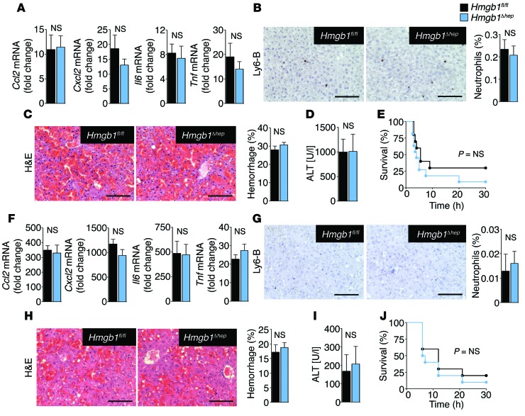
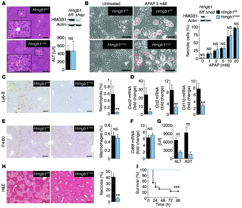
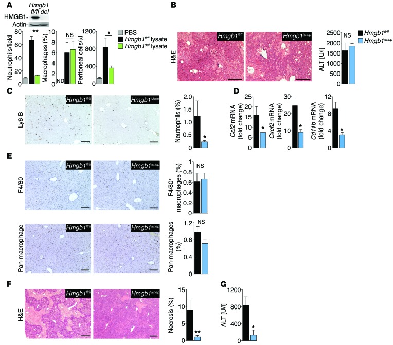
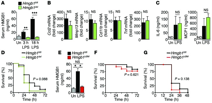
In contrast to microbially triggered inflammation, mechanisms promoting sterile inflammation remain poorly understood. Damage-associated molecular patterns (DAMPs) are considered key inducers of sterile inflammation following cell death, but the relative contribution of specific DAMPs, including high-mobility group box 1 (HMGB1), is ill defined. Due to the postnatal lethality of Hmgb1-knockout mice, the role of HMGB1 in sterile inflammation and disease processes in vivo remains controversial. Here, using conditional ablation strategies, we have demonstrated that epithelial, but not bone marrow-derived, HMGB1 is required for sterile inflammation following injury. Epithelial HMGB1, through its receptor RAGE, triggered recruitment of neutrophils, but not macrophages, toward necrosis. In clinically relevant models of necrosis, HMGB1/RAGE-induced neutrophil recruitment mediated subsequent amplification of injury, depending on the presence of neutrophil elastase. Notably, hepatocyte-specific HMGB1 ablation resulted in 100% survival following lethal acetaminophen intoxication. In contrast to necrosis, HMGB1 ablation did not alter inflammation or mortality in response to TNF- or FAS-mediated apoptosis. In LPS-induced shock, in which HMGB1 was considered a key mediator, HMGB1 ablation did not ameliorate inflammation or lethality, despite efficient reduction of HMGB1 serum levels. Our study establishes HMGB1 as a bona fide and targetable DAMP that selectively triggers a neutrophil-mediated injury amplification loop in the setting of necrosis.


Figures

Figure 6
Figure 6. RAGE-expressing neutrophils promote injury amplification after necrotic injury.
(AD) Neutrophil infiltration (A), inflammatory gene expression (B), H&E staining and necrosis (C), and serum ALT concentrations (D) in chimeric mice with WT BM (n = 8) and Ager–/– BM (n = 10) treated with acetaminophen (300 mg/kg). (EI) Chimeric mice with WT BM and Elane–/– BM (n = 12 per group) were treated with acetaminophen (300 mg/kg). Hepatic neutrophil infiltration was determined by staining for Ly6-B (E). BM reconstitution was confirmed by qPCR for Elane in spleens of BM-chimeric mice (F). Hepatic inflammatory gene expression was determined by qPCR (G). Liver injury was assessed by hepatic H&E staining and necrosis quantification (H), and serum ALT concentrations (I). *P < 0.05 and **P < 0.01 by unpaired 2-tailed t test. Scale bars: 200 μm.
Figure 5
Figure 5. RAGE, but not TLR4, mediates neutrophil recruitment following necrosis.
(A) Migration of WT, Tlr4–/–, and RAGE-deficient (RAGE encoded by Ager) neutrophils (Ager–/– neutrophils, n ≥2 separate isolations per experiment) toward WT liver lysates. GM-CSF served as a positive control. (BE) Analysis of hepatic neutrophil infiltration (B), inflammatory gene expression (C), H&E staining and necrosis quantification (D), and serum ALT concentrations (E) in WT (n = 9) and Tlr4–/– (n = 7) mice treated with acetaminophen (300 mg/kg). (FI) Analysis of hepatic neutrophil infiltration (F), inflammatory gene expression (G), H&E staining and necrosis quantification (H), and serum ALT concentrations (I) in WT (n = 10) and Ager–/– (n = 9) mice treated with acetaminophen (300 mg/kg). *P < 0.05 and **P < 0.01 by unpaired 2-tailed t test. Scale bars: 200 μm.
Figure 4
Figure 4. HMGB1 does not modulate inflammation, injury, or survival in response to FAS- or TNF-induced apoptosis.
(A and B) Hepatic inflammatory gene expression (A) and neutrophil infiltration (B) 10 hours after injection of a sublethal dose of the FAS-agonistic antibody Jo2 (0.15 μg/g) in Hmgb1fl/fl (n = 6) and Hmgb1Δhep (n = 7) mice. (C) Hepatic H&E staining and hemorrhage 5 hours after a lethal dose of Jo2 (0.5 μg/g) in Hmgb1fl/fl and Hmgb1Δhep (n = 3 per group) mice. (D and E) Serum ALT after 5 hours (D) and survival (E) in Hmgb1fl/fl (n = 9) and Hmgb1Δhep (n = 10) mice after Jo2 injection (0.5 μg/g). (FJ) Mice were injected with 700 μg/kg D-Gal and 100 μg/kg LPS. Hepatic inflammatory gene expression (F), neutrophil infiltration (G), hemorrhage (H), and serum ALT (I) 6 hours later in Hmgb1fl/fl and Hmgb1Δhep (n = 5 per group) mice. (J) Survival of Hmgb1fl/fl and Hmgb1Δhep (n = 10 per group) mice. Statistical significance assessed by 2-tailed unpaired t test (AD and FI) and Mantel-Cox log-rank test (E and J). Scale bars: 200 μm (B, C, G, and H).
Figure 3
Figure 3. HMGB1 mediates neutrophil recruitment, injury amplification, and lethality following acetaminophen-induced liver necrosis.
(A) Serum ALT and liver histology in Hmgb1fl/fl (n = 8) and Hmgb1Δhep (n = 7) mice 3 hours after injection of acetaminophen (300 mg/kg). (B) Acetaminophen-induced death in Hmgb1fl/fl and Hmgb1Δhep hepatocytes (n = 3 separate isolations per group). (CH) Hepatic neutrophil recruitment (C), inflammatory gene expression (D), macrophage numbers (E and F), serum ALT and AST (G), and liver necrosis (H) 24 hours after acetaminophen (300 mg/kg) injection in Hmgb1fl/fl (n = 8) and Hmgb1Δhep (n = 8) mice. (I) Survival after a lethal dose of acetaminophen (500 mg/kg) in Hmgb1fl/fl and Hmgb1Δhep (n = 13 per group) mice. *P < 0.05, **P < 0.01, and ***P < 0.001 by 2-tailed unpaired t test (AH) and Mantel-Cox log-rank test (I). Scale bars: 200 μm (A, C, E, and H) and 100 μm (B).
Figure 2
Figure 2. HMGB1 promotes neutrophil recruitment in vitro and in vivo.
(A) Neutrophil and macrophage migration toward Hmgb1-floxed and Hmgb1-deleted (Hmgb1del) liver extracts (induced by Mx1-Cre), determined in Boyden chambers (left 2 panels, n = 3 per group, with representative results from 3 separate isolations). Insert shows immunoblot confirming Hmgb1 deletion. Peritoneal inflammatory cell accumulation after i.p. injection of lysates from Hmgb1fl/fl (n = 7) and Hmgb1del (n = 8) livers. (BE) Hmgb1fl/fl (n = 8) and Hmgb1Δhep (n = 9) mice were subjected to warm hepatic I/R and sacrificed 6 hours later. H&E staining (B, left panel) and serum ALT (B, right panel) demonstrate similar initial injury, whereas hepatic neutrophil infiltration (C) and hepatic expression of inflammatory genes (D) differed. Numbers of hepatic macrophages determined by F4/80 staining and staining with pan-macrophage antibody in Hmgb1fl/fl (n = 8) and Hmgb1Δhep (n = 9) mice (E). (F and G) Hepatic injury 24 hours after I/R injury in Hmgb1fl/fl (n = 11) and Hmgb1Δhep (n = 10) mice, determined by H&E staining (F) and serum ALT (G). *P < 0.05 and **P < 0.01 by 1-way ANOVA followed by Tukey’s multiple comparisons test (A) and unpaired 2-tailed t test (BG). Scale bars: 200 μm.
Figure 1
Figure 1. HMGB1 does not mediate LPS-induced inflammation and lethality.
Hmgb1fl/flMx1-Creneg and Hmgb1fl/fl Mx1-Crepos mice (Hmgb1del) (AD) as well as Hmgb1fl/fl Vav1-Creneg and Hmgb1fl/fl Vav1-Crepos mice (Hmgb1ΔBM) (E and F) were treated with a lethal dose of LPS (80 mg/kg i.v.). (AC) HMGB1 serum levels (A) (n = 4 per group), hepatic inflammatory gene expression (B), and serum chemokine levels (C) were determined in Hmgb1fl/fl and Hmgb1del (n = 7 per group) mice by ELISA and qPCR, respectively. (D) Survival was determined in Hmgb1fl/fl and Hmgb1del (n = 18 per group) mice. (E and F) Hmgb1fl/fl and Hmgb1ΔBM mice were treated with LPS (30 mg/kg). HMGB1 serum levels were determined 18 hours after LPS challenge (E) (n = 4 per group). Survival was determined in Hmgb1fl/fl and Hmgb1ΔBM mice (n = 12 per group). (G) Hmgb1fl/fl and Hmgb1ΔBM mice were treated with LPS (80 mg/kg). Survival was determined in Hmgb1fl/fl (n = 15) and Hmgb1ΔBM mice (n = 10). *P < 0.05, **P < 0.01, and ***P < 0.001 by 1-way ANOVA followed by Tukey’s multiple comparisons test (A, C, and E), unpaired 2-tailed t test (B), and Mantel-Cox log-rank test (D, F, and G), respectively. Un, untreated; ND, nondetectable; NS, nonsignificant.

References

    1. Kono H, Rock KL. How dying cells alert the immune system to danger. Nat Rev Immunol. 2008;8(4):279–289. doi: 10.1038/nri2215. - DOI - PMC - PubMed
    1. Matzinger P. The danger model: a renewed sense of self. Science. 2002;296(5566):301–305. doi: 10.1126/science.1071059. - DOI - PubMed
    1. Scaffidi P, Misteli T, Bianchi ME. Release of chromatin protein HMGB1 by necrotic cells triggers inflammation. Nature. 2002;418(6894):191–195. doi: 10.1038/nature00858. - DOI - PubMed
    1. Lotze MT, Tracey KJ. High-mobility group box 1 protein (HMGB1): nuclear weapon in the immune arsenal. Nat Rev Immunol. 2005;5(4):331–342. doi: 10.1038/nri1594. - DOI - PubMed
    1. Zitvogel L, Kepp O, Kroemer G. Decoding cell death signals in inflammation and immunity. Cell. 2010;140(6):798–804. doi: 10.1016/j.cell.2010.02.015. - DOI - PubMed

Publication types

MeSH terms