. 2015 Jan 6;13(1):e1002036.
doi: 10.1371/journal.pbio.1002036. eCollection 2015 Jan.

Distinct brain systems mediate the effects of nociceptive input and self-regulation on pain

Affiliations

Distinct brain systems mediate the effects of nociceptive input and self-regulation on pain

Choong-Wan Woo et al. PLoS Biol. .

Abstract

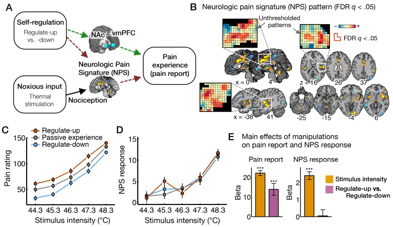
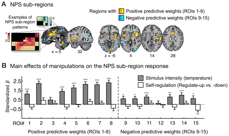
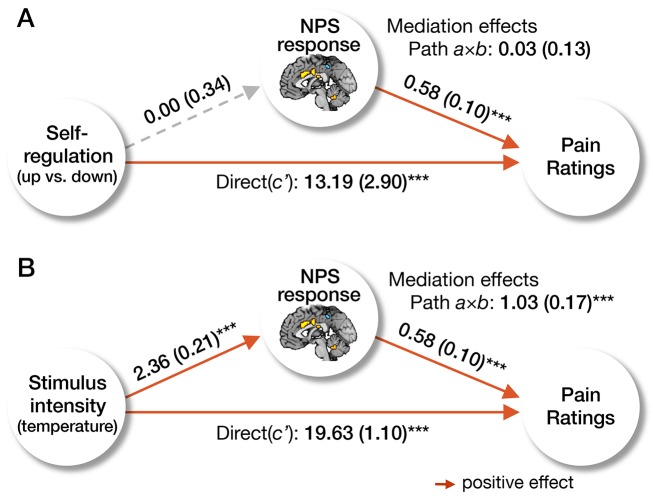
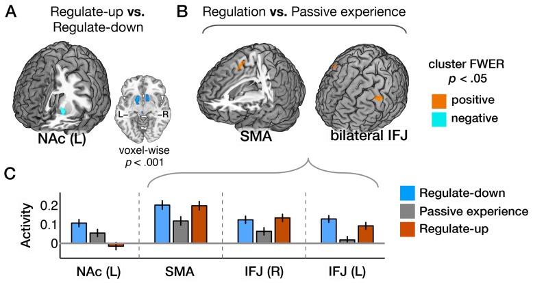
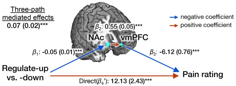
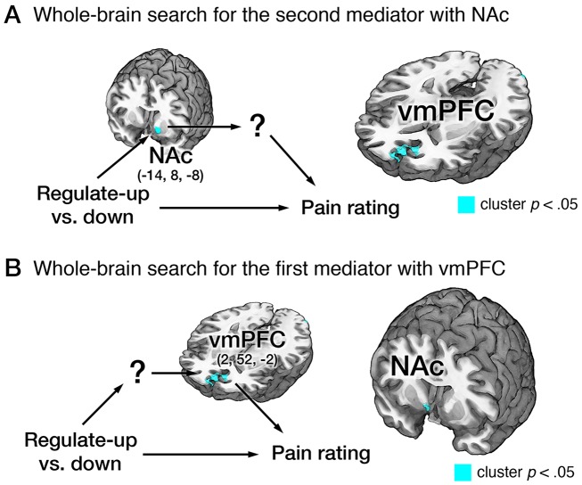
Cognitive self-regulation can strongly modulate pain and emotion. However, it is unclear whether self-regulation primarily influences primary nociceptive and affective processes or evaluative ones. In this study, participants engaged in self-regulation to increase or decrease pain while experiencing multiple levels of painful heat during functional magnetic resonance imaging (fMRI) imaging. Both heat intensity and self-regulation strongly influenced reported pain, but they did so via two distinct brain pathways. The effects of stimulus intensity were mediated by the neurologic pain signature (NPS), an a priori distributed brain network shown to predict physical pain with over 90% sensitivity and specificity across four studies. Self-regulation did not influence NPS responses; instead, its effects were mediated through functional connections between the nucleus accumbens and ventromedial prefrontal cortex. This pathway was unresponsive to noxious input, and has been broadly implicated in valuation, emotional appraisal, and functional outcomes in pain and other types of affective processes. These findings provide evidence that pain reports are associated with two dissociable functional systems: nociceptive/affective aspects mediated by the NPS, and evaluative/functional aspects mediated by a fronto-striatal system.


Conflict of interest statement

The authors have declared that no competing interests exist.

Figures

Figure 1
Figure 1. The effects of nociceptive input and cognitive self-regulation on reported pain and brain activity.
(A) Hypotheses about the effects of cognitive regulation on pain-related brain processes. The effects of cognitive self-regulation on pain could be mediated by the NPS (hypothesis A, dark-brown dashed lines) or other brain systems, particularly a fronto-striatal pathway connecting vmPFC and NAc (hypothesis B, green dashed lines). (B) The NPS pattern, an a priori distributed pattern of fMRI signal that is sensitive and specific to physical pain . Here, we present the thresholded pattern map (q<0.05, false discovery rate [FDR]) for display only, but all voxels within NPS were used in analyses. Some examples of unthresholded patterns are also presented in the insets; small squares indicate voxel weights, and black squares indicate empty voxels located outside of the NPS mask. (C,D) Pain ratings and NPS response as a function of stimulus intensity and regulation conditions. Because the highest level of heat intensity, 49.3°C, was not used in the self-regulation runs, we displayed results only for five levels of stimulus intensity. However all levels of heat intensity were included in analyses. NPS response values, which indicate the strength of expression of the signature pattern, are calculated by taking the dot-product of the NPS pattern weights and activation maps for each single trial. Error bars represent within-subject standard errors of the mean (SEM). The numerical data used to generate the plots can be found in Table S1. (E) The main effects of manipulations (stimulus intensity and regulate-up versus -down) on pain ratings and NPS response. Beta values (y-axis) represent regression coefficients from a multilevel generalized linear model. Error bars represent SEM. *** p<0.001, two-tailed.
Figure 2
Figure 2. The effects of stimulus intensity and self-regulation on separate sub-regions within the neurologic pain signature.
(A) NPS sub-regions: ROIs 1–8 are regions with positive NPS weights, and ROIs 9–15 are regions with negative NPS weights. We obtained these regions from the FDR (q<0.05) thresholded NPS map (see Figure 1B) smoothed with a 0.5 mm Gaussian kernel. Some examples of weight patterns within the NPS sub-regions are presented in the insets; small squares indicate voxel weights, and black squares indicate empty voxels located outside of the ROIs. (B) The main effects of stimulus intensity and self-regulation on the NPS response within sub-regions. y-Axis represents standardized regression coefficients from multilevel generalized linear models with stimulus intensity and self-regulation (regulate-up versus -down) as predictors and NPS response as dependent variables. Error bars represent standard errors of the mean (SEM). The data used to generate the plots can be found in Table S2. ***p<0.001; **p<0.01; *p<0.05, two-tailed.
Figure 3
Figure 3. Mediation of the neurologic pain signature.
We present the results of the multilevel mediation analyses with the NPS response as a mediator. In (A), cognitive self-regulation (regulate-up versus -down instructions) was entered as a predictor, and in (B), stimulus intensity (i.e., temperature) was entered as a predictor. In both models, pain report was an outcome, and the other manipulation (e.g., stimulus intensity for model A, and self-regulation for model B) was entered as a covariate. The results showed that the NPS response mediated the effects of stimulus intensity on pain, but did not mediate the effects of cognitive regulation on pain. The paths (path a, b, and c′) and mediation effects (path a×b) are labeled with path coefficients, and their standard errors are shown in parenthesis. The gray dashed line indicates a non-significant path. ***p<0.001, two-tailed.
Figure 4
Figure 4. Brain activity induced by self-regulation.
(A) Activity in left NAc was associated with regulate-up versus regulate-down instructions (at p<0.05, FWER corrected based on cluster extent, with a primary threshold of p<0.0005, k>84). Bilateral activations were found at a lower threshold (voxel-wise p<0.001). (B) Activity in the supplementary motor area (SMA) and bilateral inferior frontal junction (IFJ) were associated with regulation versus passive experience instructions. (C) Bar plots of the averaged activity (y-axis) within the corresponding brain regions for conditions (x-axis). Error bars represent within-subject standard errors of the mean (SEM). The data used to generate the plots can be found in Table S4.
Figure 5
Figure 5. Multilevel three-path mediation analysis with two a priori regions-of-interest.
A priori ROIs include the NAc (MNI: 10, 12, −8) and the vmPFC (MNI: 2, 52, −2) from Baliki and colleagues . Stimulus intensity and the NPS response were included as covariates. The paths are labeled with path coefficients, and standard errors are shown in parentheses (for more details about three-path mediation analyses, see Materials and Methods). *** p<0.001, two-tailed.
Figure 6
Figure 6. Whole-brain three-path mediation analysis results.
(A) Whole-brain three-path mediation analysis with the NAc (MNI: −14, 8, −8), which is from the GLM results shown in Figure 4A, as the first mediator. VMPFC (MNI: 2, 52, −2) was the only significant second brain mediator from the whole-brain search (p<0.05, FWER corrected based on cluster extent, with a primary threshold of p<0.001). (B) Whole-brain three-path mediation analysis with vmPFC (MNI: 2, 52, −2), which is from the results shown in Figure 6A. Right NAc (MNI: 8, 8, −6) was the only significant first brain mediator from the whole-brain search. For more details of path coefficients, see Table S5. *** p<0.001, two-tailed.

Comment in

  • Pain: a distributed brain information network?
    Mano H, Seymour B. Mano H, et al. PLoS Biol. 2015 Jan 6;13(1):e1002037. doi: 10.1371/journal.pbio.1002037. eCollection 2015 Jan. PLoS Biol. 2015. PMID: 25562782 Free PMC article.
  • Pain: reappraising pain.
    Bray N. Bray N. Nat Rev Neurosci. 2015 Mar;16(3):124. doi: 10.1038/nrn3919. Epub 2015 Jan 29. Nat Rev Neurosci. 2015. PMID: 25630993 No abstract available.
  • Towards a taxonomy of pain modulations.
    Ploner M, Bingel U, Wiech K. Ploner M, et al. Trends Cogn Sci. 2015 Apr;19(4):180-2. doi: 10.1016/j.tics.2015.02.007. Epub 2015 Mar 4. Trends Cogn Sci. 2015. PMID: 25745857

References

    1. Gross JJ, Munoz RF (1995) Emotion regulation and mental-health. Clin Psychol-Sci Pr 2: 151–164.
    1. Ochsner KN, Silvers JA, Buhle JT (2012) Functional imaging studies of emotion regulation: a synthetic review and evolving model of the cognitive control of emotion. Ann N Y Acad Sci 1251: E1–E24. - PMC - PubMed
    1. Buhle JT, Silvers JA, Wager TD, Lopez R, Onyemekwu C, et al. (2013) Cognitive reappraisal of emotion: a meta-analysis of human neuroimaging studies. Cereb Cortex 24: 2981–2990. - PMC - PubMed
    1. Ochsner KN, Bunge SA, Gross JJ, Gabrieli JD (2002) Rethinking feelings: an FMRI study of the cognitive regulation of emotion. J Cogn Neurosci 14: 1215–1229. - PubMed
    1. Goldin PR, McRae K, Ramel W, Gross JJ (2008) The neural bases of emotion regulation: reappraisal and suppression of negative emotion. Biol Psychiatry 63: 577–586. - PMC - PubMed

Publication types

LinkOut - more resources