

Debilitating lung disease among surface coal miners with no underground mining tenure
- PMID: 25563541
- PMCID: PMC4626873
- DOI: 10.1097/JOM.0000000000000302
Debilitating lung disease among surface coal miners with no underground mining tenure
Abstract
Objective: To characterize exposure histories and respiratory disease among surface coal miners identified with progressive massive fibrosis from a 2010 to 2011 pneumoconiosis survey.
Methods: Job history, tenure, and radiograph interpretations were verified. Previous radiographs were reviewed when available. Telephone follow-up sought additional work and medical history information.
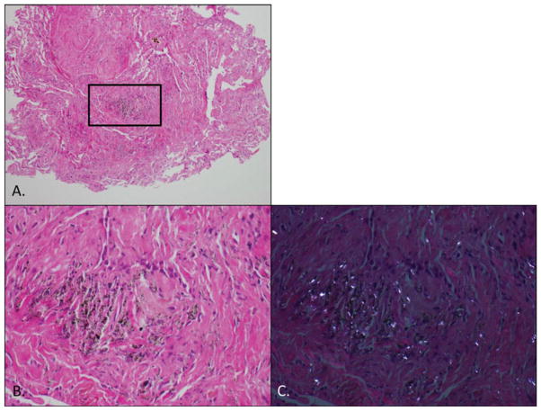
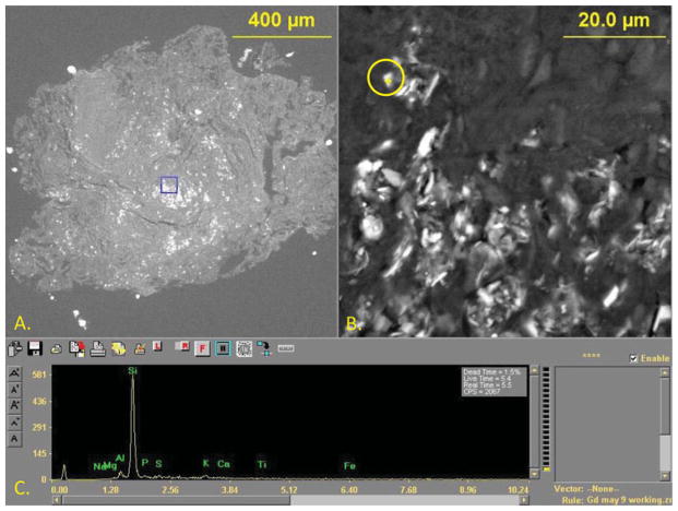
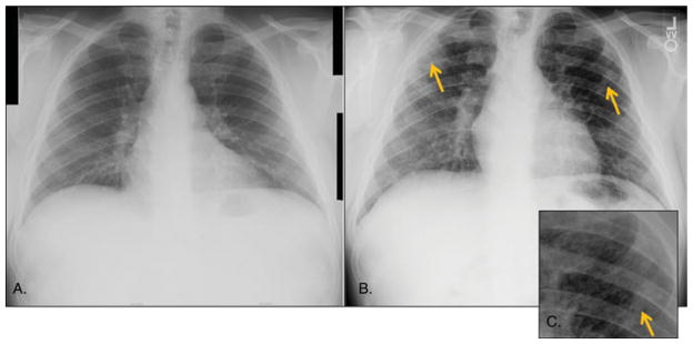
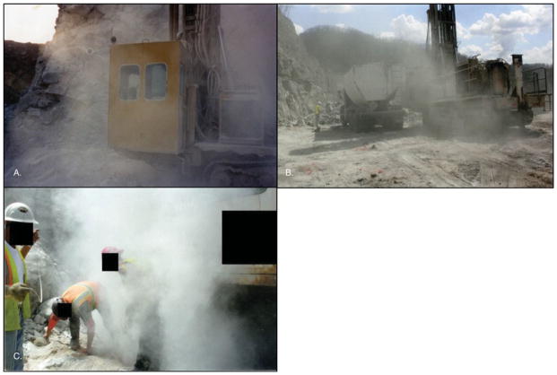
Results: Among eight miners who worked as drill operators or blasters for most of their tenure (median, 35.5 years), two reported poor dust control practices, working in visible dust clouds as recently as 2012. Chest radiographs progressed to progressive massive fibrosis in as few as 11 years. One miner's lung biopsy demonstrated fibrosis and interstitial accumulation of macrophages containing abundant silica, aluminum silicate, and titanium dust particles.
Conclusions: Overexposure to respirable silica resulted in progressive massive fibrosis among current surface coal miners with no underground mining tenure. Inadequate dust control during drilling/blasting is likely an important etiologic factor.
Conflict of interest statement
The authors declare no conflicts of interest.
Figures
References
-
- U.S. Department of Labor; Mine Safety and Health Administration (MSHA) [Accessed May 2014];Lowering Miners’ Exposure to Respirable Coal Mine Dust, Including Continuous Personal Dust Monitors. Available at https://s3.amazonaws.com/public-inspection.federalregister.gov/2014-0908....
-
- Parker J, Lapp N, Banks D. Surface coal mine drillers and silicosis: the ten year West Virginia experience. Am Rev Respir Dis. 1989;139:A490.
-
- Amandus HE, Hanke W, Kullman G, et al. A re-evaluation of radiological evidence from a study of US strip coal miners. Arch Environ Health. 1984;39:346–351. - PubMed
-
- Amandus HE, Petersen MR, Richards TB. Health status of anthracite surface coal miners. Arch Environ Health. 1989;44:75–81. - PubMed
-
- Centers for Disease Control and Prevention. Silicosis screening in surface coal miners—Pennsylvania, 1996–1997. MMWR Morb Mortal Wkly Rep. 2000;49:612–615. - PubMed
Publication types
MeSH terms
Grants and funding
LinkOut - more resources
Full Text Sources
Medical
