

Vascular imaging abnormalities and cognition: mediation by cortical volume in nondemented individuals: atherosclerosis risk in communities-neurocognitive study
- PMID: 25563642
- PMCID: PMC4308430
- DOI: 10.1161/STROKEAHA.114.007847
Vascular imaging abnormalities and cognition: mediation by cortical volume in nondemented individuals: atherosclerosis risk in communities-neurocognitive study
Abstract
Background and purpose: The relationships between cerebrovascular lesions visible on imaging and cognition are complex. We explored the possibility that the cerebral cortical volume mediated these relationships.
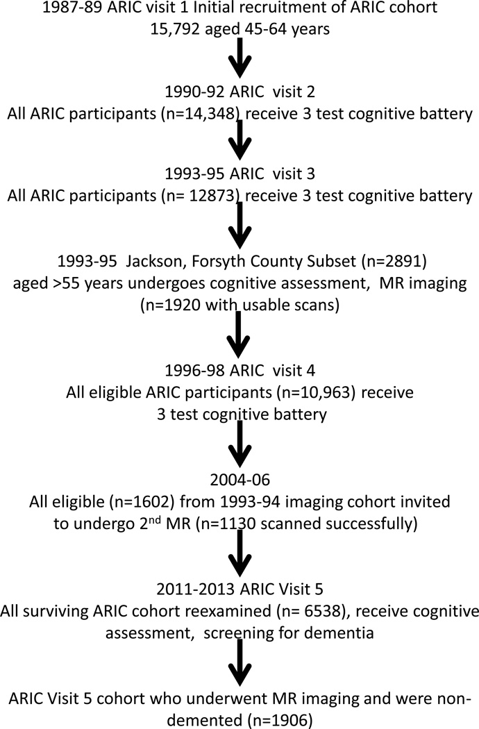
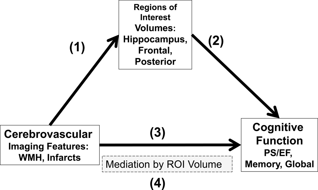
Methods: Total of 1906 nondemented participants (59% women; 25% African-American; mean age, 76.6 years) in the Atherosclerosis Risk in Communities (ARIC) study underwent cognitive assessments, risk factor assessments, and quantitative MRI for white matter hyperintensities (WMH) and infarcts. The Freesurfer imaging analysis pipeline was used to determine regional cerebral volumes. We examined the associations of cognitive domain outcomes with cerebral volumes (hippocampus and separate groups of posterior and frontal cortical regions of interest) and cerebrovascular imaging features (presence of large or small cortical/subcortical infarcts and WMH volume). We performed mediation pathway analyses to assess the hypothesis that hippocampal and cortical volumes mediated the associations between cerebrovascular imaging features and cognition.
Results: In unmediated analyses, WMH and infarcts were both associated with worse psychomotor speed/executive function. In mediation analyses, WMH and infarct associations on psychomotor speed/executive function were significantly attenuated, but not abolished, by the inclusion of the posterior cortical regions of interest volume in the models, and the infarcts on psychomotor speed/executive function association were attenuated, but not abolished, by inclusion of the frontal cortical regions of interest volume.
Conclusions: Both WMH and infarcts were associated with cortical volume, and both lesions were also associated with cognitive performance, implying shared pathophysiological mechanisms. Although cross-sectional, our findings suggest that WMH and infarcts could be proxies for clinically covert processes that directly damage cortical regions. Microinfarcts are 1 candidate for such a clinically covert process.
Keywords: cerebral infarction; cerebral small vessel diseases; cognition; magnetic resonance imaging.
© 2015 American Heart Association, Inc.
Figures
References
-
- Blessed G, Tomlinson BE, Roth M. The association between quantitative measures of dementia and of senile change in the cerebral grey matter of elderly subjects. Br J Psychiatry. 1968;114:797–811. - PubMed
-
- Schneider JA, Arvanitakis Z, Bang W, Bennett DA. Mixed brain pathologies account for most dementia cases in community-dwelling older persons. Neurology. 2007;69:2197–2204. - PubMed
-
- Knopman D, Parisi JE, Boeve BF, Rocca WA, Cha RH, Apaydin H, et al. Vascular Dementia in a Population-based autopsy study. Arch Neurol. 2003;60:569–576. - PubMed
Publication types
MeSH terms
Grants and funding
LinkOut - more resources
Full Text Sources
Medical
