. 2015 Mar;11(1):139-53.
doi: 10.1007/s11302-014-9442-3. Epub 2015 Jan 8.

Shear stress modulates endothelial KLF2 through activation of P2X4

Affiliations

Shear stress modulates endothelial KLF2 through activation of P2X4

R Sathanoori et al. Purinergic Signal. 2015 Mar.

Abstract

Vascular endothelial cells that are in direct contact with blood flow are exposed to fluid shear stress and regulate vascular homeostasis. Studies report endothelial cells to release ATP in response to shear stress that in turn modulates cellular functions via P2 receptors with P2X4 mediating shear stress-induced calcium signaling and vasodilation. A recent study shows that a loss-of-function polymorphism in the human P2X4 resulting in a Tyr315>Cys variant is associated with increased pulse pressure and impaired endothelial vasodilation. Although the importance of shear stress-induced Krüppel-like factor 2 (KLF2) expression in atheroprotection is well studied, whether ATP regulates KLF2 remains unanswered and is the objective of this study. Using an in vitro model, we show that in human umbilical vein endothelial cells (HUVECs), apyrase decreased shear stress-induced KLF2, KLF4, and NOS3 expression but not that of NFE2L2. Exposure of HUVECs either to shear stress or ATPγS under static conditions increased KLF2 in a P2X4-dependent manner as was evident with both the receptor antagonist and siRNA knockdown. Furthermore, transient transfection of static cultures of human endothelial cells with the Tyr315>Cys mutant P2X4 construct blocked ATP-induced KLF2 expression. Also, P2X4 mediated the shear stress-induced phosphorylation of extracellular regulated kinase-5, a known regulator of KLF2. This study demonstrates a major physiological finding that the shear-induced effects on endothelial KLF2 axis are in part dependent on ATP release and P2X4, a previously unidentified mechanism.


Figures

Fig. 1
Fig. 1
HUVECs express P2X4 receptors. Flow cytometric analysis of HUVECs using an anti-P2X4 antibody recognizing an extracellular epitope shows these cells to express P2X4 receptors (a; in red) and pre-adsorption of the antibody with the corresponding antigen shows antibody specificity (b; in blue). Unstained population (in black) and isotype control (in green) are also shown in both the histograms. A representative immunoblot (c) probed with an anti-P2X4 antibody recognizing an intracellular epitope shows P2X4 receptor at approximately 60 kDa. The lane assignments are as follows: lane 1 THP-1 cell lysate probed with P2X4 antibody pre-adsorbed with P2X4 peptide, lane 2 THP-1 cell lysate, lane 3 marker lane showing bands at 50 and 75 kDa, and lane 4 HUVEC cell lysate
Fig. 2
Fig. 2
Validation of the in vitro orbital shaker model of shear stress. Representative bright-field images (×10 objective) show morphology (a) of HUVECs exposed to either static (ST) (left panel) or cells cultured in the peripheral area of the IVF dish using an orbital shaker to shear stress (SS) conditions for 48 h (0.1 Pa or 1 dyne/cm2; middle panel and 1 Pa or 10 dynes/cm2; right panel). qRT-PCR analysis reveals altered P2X4 (b) and KLF2 (c) at 1 Pa but not 0.1 Pa shear stress. Time course of cells cultured under shear stress conditions (1 Pa) for 3, 6, 12, 24, and 48 h show cell alignment as early as 3 h (d) and qRT-PCR analysis show time-dependent decrease of P2X4 (e) as well as an increase in KLF2 (f) transcript levels normalized to the geometric mean of housekeeping genes (HKG). Quantitation of Westerns along with representative immunoblots show shear stress to decrease P2X4 (g) and increase KLF2 (h) protein as normalized to the loading control (β-tubulin). Representative epifluorescent images (×20 objective) (i) of HUVECs subjected to 6 h (a and b) and 24 h (c and d) static and shear stress conditions (1 Pa) show an increase in KLF2 (Northern Lights™-567). Actin cytoskeleton and nucleus were stained with ActinGreen™-488 and NucBlue®, respectively. White arrows in a, d, and i indicate the direction of flow. N = 3–6 experiments each in replicates; *p ≤ 0.05. Scale bar, 20 μM
Fig. 3
Fig. 3
Apyrase decreases shear stress-induced KLF2, NOS3, and KLF4, but not NFE2L2 expression. HUVECs were cultured for 6, 24, and 48 h (a) under static (no shear stress) or shear stress conditions (1 Pa) in the presence or absence of 2 U/ml of apyrase, an ATP hydrolyzing enzyme with the line graph depicting fold change in KLF2 mRNA (a). Fold change in KLF4 (b), NOS3 (c), and NFE2L2 (d) mRNA at 48 h with all transcripts normalized to the geometric mean of the housekeeping genes (HKG). N = 3–4 experiments in replicates; *p ≤ 0.05
Fig. 4
Fig. 4
ATPγS induces KLF2 in a P2X4-dependent manner. HUVECs were cultured for 3 h under static conditions in the presence or absence of 200 μmol/L ATPγS, 100 μmol/L UTP, and 10 μmol/L 2MeSADP with the bar graph (a) depicting fold change in KLF2 mRNA normalized to the geometric mean of the housekeeping genes (HKG). Static HUVEC cultures were exposed to 200 μmol/L ATPγS for 3 h in the presence or absence of 1 μmol/L PSB-12253. qRT-PCR analysis (b) shows the effect of PSB-12253 on KLF2 mRNA. Representative epifluorescent images (×40 objective) (c) of KLF2 immunostaining (Northern Lights™-567), actin cytoskeleton (ActinGreen™-488), and nucleus (NucBlue®) in cultures exposed to PSB-12253. Panels ad represent KLF2 and (a’d’) merged images of KLF2, actin, and DAPI staining. N = 2–3 experiments in replicates; *p ≤ 0.05. Scale bar, 20 μM
Fig. 5
Fig. 5
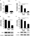
Validation of siRNA knockdown of P2X4 receptors. Validation of the knockdown of P2X4 48 h post-transfection in static (a and b) and shear stress conditions (c and d) by qRT-PCR (a and c) and Western (b and d) with representative immunoblots for P2X4 (60 kDa) and the loading control, β-tubulin (50 kDa). N = 4–6 experiments each in replicates; *p ≤ 0.05; HKG housekeeping genes
Fig. 6
Fig. 6
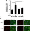
siRNA knockdown of P2X4 inhibits ATPγS-induced KLF2. Static HUVEC cultures transfected with negative control and P2X4 siRNA for 48–72 h were exposed to 200 μmol/L ATPγS for 3 h. qRT-PCR analysis (a) shows the effect of siRNA knockdown (48 h) of P2X4 on KLF2 mRNA normalized to the geometric mean of the housekeeping genes (HKG). Representative epifluorescent images (×40 objective) (b) of KLF2 immunostaining (Northern Lights™-567), actin cytoskeleton (ActinGreen™-488), and nucleus (NucBlue®) in cultures with siRNA knockdown (72 h) of P2X4. Panels (a–d) represent KLF2 and (a’–d’) merged images of KLF2, actin, and DAPI staining. N = 3 experiments in replicates; *p ≤ 0.05. Scale bar, 20 μM
Fig. 7
Fig. 7
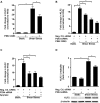
Selective antagonist and siRNA knockdown of P2X4 receptors reduce shear stress-induced KLF2. HUVECs were cultured for 6 h under static or shear stress conditions (1 Pa) in the presence or absence of the P2X4 receptor antagonist, PSB-12253 (1 μmol/L) (a and b) or transiently transfected with siRNA (bd). Graphs depict fold change in transcript levels of KLF2 (a) in response to PSB-12253. Knocking down of P2X4 results in decreased levels of shear stress-induced KLF2 mRNA (b and c) with no additive effect of the antagonist (b) or apyrase (c) after siRNA knockdown. Transcript levels were normalized to the geometric mean of the housekeeping genes (HKG). In addition, P2X4 siRNA inhibits KLF2 protein (d) with a representative immunoblot showing levels of KLF2 and the loading control β-tubulin after siRNA knockdown of P2X4. Fold changes are relative to cells transfected with the negative control siRNA. N = 3–6 experiments in replicates; *p ≤ 0.05
Fig. 8
Fig. 8
Ivermectin increases ATPγS-induced KLF2 at 6 h. Static HUVEC cultures were exposed to 200 μmol/L of ATPγS in the presence or absence of 3 μmol/L ivermectin for 6 h. qRT-PCR analysis shows a significant increase in ATPγS-induced KLF2 transcript levels only in the presence of ivermectin (a) with no synergistic effect after siRNA knockdown (b) of P2X4. N = 3 experiments in replicates; *p ≤ 0.05; HKG housekeeping genes
Fig. 9
Fig. 9
Y315C mutant P2X4 inhibits ATPγS-induced gene expression. Static HUVEC cultures transiently transfected for 48 h with WT and Y315C mutant P2X4 constructs were exposed to 200 μmol/L ATPγS for 3 h. qRT-PCR analysis shows the effect of the SNP on KLF2 (a), NOS3 (b), THBD (c), KLF4 (d), and NFE2L2 (e) mRNA. N = 2 experiments in replicates; *p ≤ 0.05
Fig. 10
Fig. 10
Shear stress induced ERK5 phosphorylation in a P2X4-dependent receptor. HUVECs transiently transfected for 48 h with P2X4 siRNA were cultured for 6 h under static or shear stress (1 Pa) conditions. Graph depict fold difference in shear stress-induced phosphorylated ERK5 (Thr218/Tyr220) normalized to total ERK5, which is dependent on P2X4 with a representative immunoblot showing levels of phosphorylated ERK5 and total ERK5. Fold difference is relative to cells transfected with the negative control siRNA. N = 6 experiments in replicates; *p ≤ 0.05

Similar articles

Cited by

References

    1. Gimbrone MA, Jr, Garcia-Cardena G. Vascular endothelium, hemodynamics, and the pathobiology of atherosclerosis. Cardiovasc Pathol. 2013;22:9–15. doi: 10.1016/j.carpath.2012.06.006. - DOI - PMC - PubMed
    1. Davies PF. Hemodynamic shear stress and the endothelium in cardiovascular pathophysiology. Nature clinical practice. Cardiovasc Med. 2009;6:16–26. - PMC - PubMed
    1. Gimbrone MA, Jr, Topper JN, Nagel T, Anderson KR, Garcia-Cardena G. Endothelial dysfunction, hemodynamic forces, and atherogenesis. Ann NY Acad Sci. 2000;902:230–239. doi: 10.1111/j.1749-6632.2000.tb06318.x. - DOI - PubMed
    1. Lin Z, Natesan V, Shi H, Dong F, Kawanami D, Mahabeleshwar GH, Atkins GB, Nayak L, Cui Y, Finigan JH, Jain MK. Kruppel-like factor 2 regulates endothelial barrier function. Arterioscler Thromb Vasc Biol. 2010;30:1952–1959. doi: 10.1161/ATVBAHA.110.211474. - DOI - PMC - PubMed
    1. Takada Y, Shinkai F, Kondo S, Yamamoto S, Tsuboi H, Korenaga R, Ando J. Fluid shear stress increases the expression of thrombomodulin by cultured human endothelial cells. Biochem Biophys Res Commun. 1994;205:1345–1352. doi: 10.1006/bbrc.1994.2813. - DOI - PubMed

Publication types

MeSH terms

LinkOut - more resources