Telemedicine for detecting diabetic retinopathy: a systematic review and meta-analysis
- PMID: 25563767
- PMCID: PMC4453504
- DOI: 10.1136/bjophthalmol-2014-305631
Telemedicine for detecting diabetic retinopathy: a systematic review and meta-analysis
Abstract
Objective: To determine the diagnostic accuracy of telemedicine in various clinical levels of diabetic retinopathy (DR) and diabetic macular oedema (DME).
Methods: PubMed, EMBASE and Cochrane databases were searched for telemedicine and DR. The methodological quality of included studies was evaluated using the Quality Assessment for Diagnostic Accuracy Studies (QUADAS-2). Measures of sensitivity, specificity and other variables were pooled using a random effects model. Summary receiver operating characteristic curves were used to estimate overall test performance. Meta-regression and subgroup analyses were used to identify sources of heterogeneity. Publication bias was evaluated using Stata V.12.0.
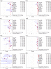
Results: Twenty articles involving 1960 participants were included. Pooled sensitivity of telemedicine exceeded 80% in detecting the absence of DR, low- or high-risk proliferative diabetic retinopathy (PDR), it exceeded 70% in detecting mild or moderate non-proliferative diabetic retinopathy (NPDR), DME and clinically significant macular oedema (CSME) and was 53% (95% CI 45% to 62%) in detecting severe NPDR. Pooled specificity of telemedicine exceeded 90%, except in the detection of mild NPDR which reached 89% (95% CI 88% to 91%). Diagnostic accuracy was higher with digital images obtained through mydriasis than through non-mydriasis, and was highest when a wide angle (100-200°) was used compared with a narrower angle (45-60°, 30° or 35°) in detecting the absence of DR and the presence of mild NPDR. No potential publication bias was detected.
Conclusions: The diagnostic accuracy of telemedicine using digital imaging in DR is overall high. It can be used widely for DR screening. Telemedicine based on the digital imaging technique that combines mydriasis with a wide angle field (100-200°) is the best choice in detecting the absence of DR and the presence of mild NPDR.
Keywords: Diagnostic tests/Investigation; Retina; Telemedicine.
Published by the BMJ Publishing Group Limited. For permission to use (where not already granted under a licence) please go to http://group.bmj.com/group/rights-licensing/permissions.
Figures


References
-
- International Diabetes Federation. Diabetes atlas. 6th edn http://www.idf.org/diabetesatlas
-
- [No authors listed] Grading diabetic retinopathy from stereoscopic color fundus photographs—an extension of the modified Airlie House classification. ETDRS report number 10. Early Treatment Diabetic Retinopathy Study Research Group. Ophthalmology 1991;98(5 Suppl):786–806. 10.1016/S0161-6420(13)38012-9 - DOI - PubMed
Publication types
MeSH terms
LinkOut - more resources
Full Text Sources
Other Literature Sources
Medical
