

doi: 10.1039/c4cc09412c.
Active site-directed proteomic probes for adenylation domains in nonribosomal peptide synthetases
Affiliations
- PMID: 25563804
- PMCID: PMC4795001
- DOI: 10.1039/c4cc09412c
Item in Clipboard
Active site-directed proteomic probes for adenylation domains in nonribosomal peptide synthetases
Chem Commun (Camb).
.
Abstract
We describe a general strategy for selective chemical labeling of individual adenylation (A) domains in nonribosomal peptide synthetases (NRPSs) using active site-directed proteomic probes coupled to the 5'-O-N-(aminoacyl)sulfamoyladenosine (AMS) scaffold with a clickable benzophenone functionality. These proteomic tools can greatly facilitate the molecular identification, functional characterization, and profiling of virtually any kind of A domains of NRPS enzymes in complex biological systems.
Figures
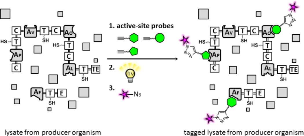
Methods for proteomic analysis of A domains in NRPS biosynthetic enzymes using active site-directed proteomic probes for A domains. Modules are comprised of thiolation (T), adenylation (A) [Af : l -Phe; Ap : l -Pro; Av : l -Val; Ao : l -Orn specific A domains[, epimerization (E), condensation (C), and thioesterase (TE) domains. In a gel-based analysis, recombinant NRPS enzymes or proteomes treated with individual probes are incubated with a rhodamine (Rh)-azide reporter tag under click chemistry (CC) conditions and separated by SDS-PAGE, and labeled A domains in NRPS enzymes are visualized by in-gel fluorescence imaging.
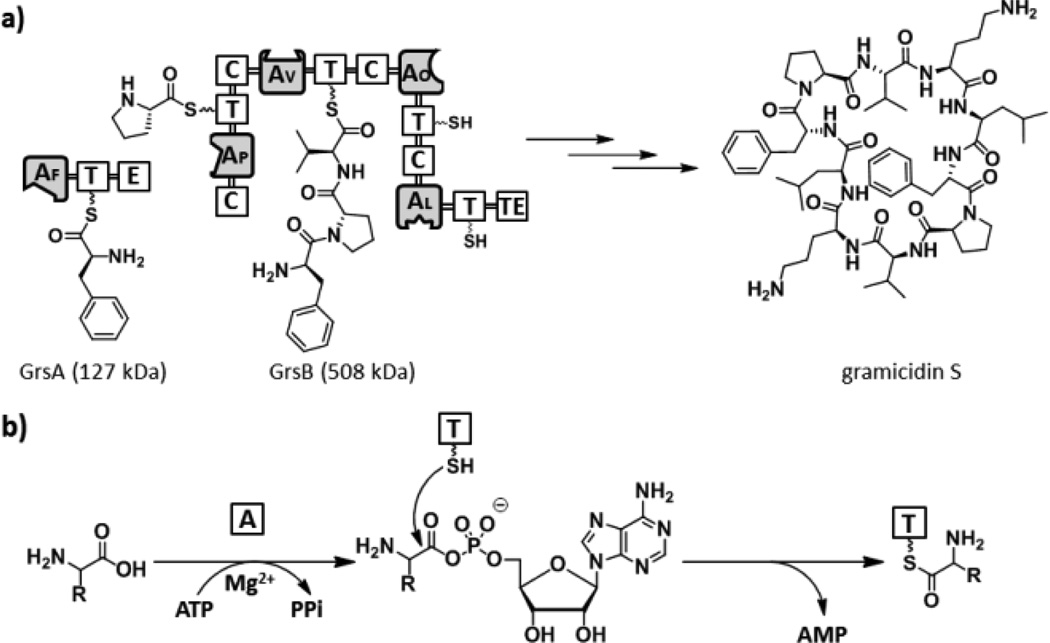
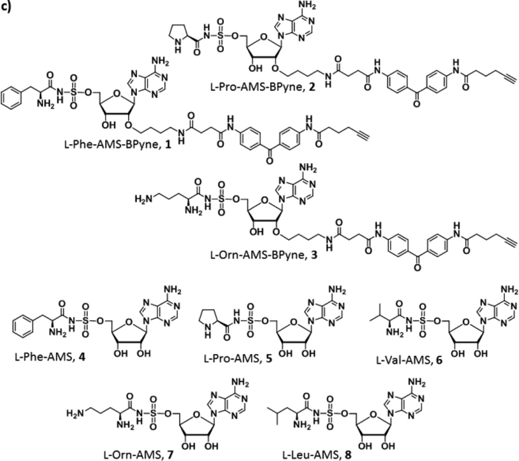
(a) Biosynthetic pathway of the gramicidine S. GrsA is composed of A1 (l -Phe), T, and E domains. In contrast, GrsB consists of four NRPS modules, C-A2 (l -Pro)-T-C-A3 (l -Val)-T-C-A4 (l -Orn)-T-C-A5 (l -Leu)-T-TE. (b) The adenylation reaction in NRPS. (c) Structures of active site-directed proteomic probes and inhibitors for A domains described in this study.
(a) Biosynthetic pathway of the gramicidine S. GrsA is composed of A1 (l -Phe), T, and E domains. In contrast, GrsB consists of four NRPS modules, C-A2 (l -Pro)-T-C-A3 (l -Val)-T-C-A4 (l -Orn)-T-C-A5 (l -Leu)-T-TE. (b) The adenylation reaction in NRPS. (c) Structures of active site-directed proteomic probes and inhibitors for A domains described in this study.
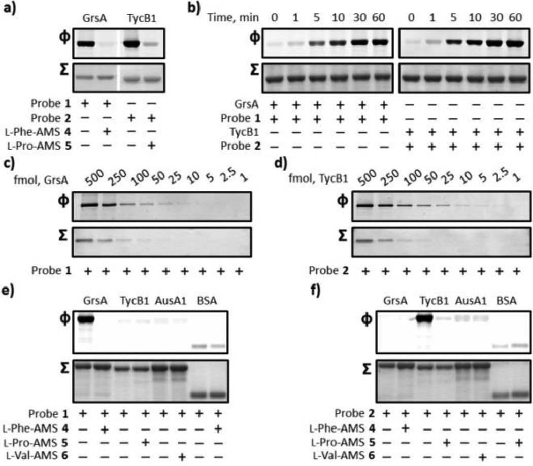
Labeling of recombinant holo-GrsA and holo-TycB1 with probes 1 and 2. (a) Labeling of GrsA and TycB1 and competitive inhibition studies with excess inhibitors 4 and 5. GrsA (1 µM) and TycB1 (1 µM) were individually pre-incubated in either the absence or presence of 100 µM of inhibitors 4 and 5 and treated with 1 µM of the individual probes 1 and 2. (b) Ultraviolet photolysis time course studies of the labeling of GrsA with probe 1 (left) and TycB1 with probe 2 (right). SDS-PAGE analysis denoting the labeling of 1 µM of GrsA and TycB1 with 1 µM probes 1 and 2, respectively. (c, d) Limit of detection of GrsA and TycB1 labeling. GrsA (0.125–62.5 nM) and TycB1 (0.125–62.5 nM) were individually incubated with probes 1 and 2. (e, f) Labeling specificity of probes 1 and 2. GrsA (1 µM), TycB1 (1 µM), AusA1 (1 µM), and BSA (1 µM) were individually treated with 1 µM of probes 1 and 2 in either the absence or presence of 100 µM of the inhibitors 4, 5, and 6. For each panel, Φ depicts the fluorescence observed with λex = 532 nm and λem = 580 nm, and Σ displays the total protein content by staining with Coomassie Blue (a, b, e, and f) or a silver staining method (c and d). Full gels (Figures S3) and experimental procedures are provided in the SI.
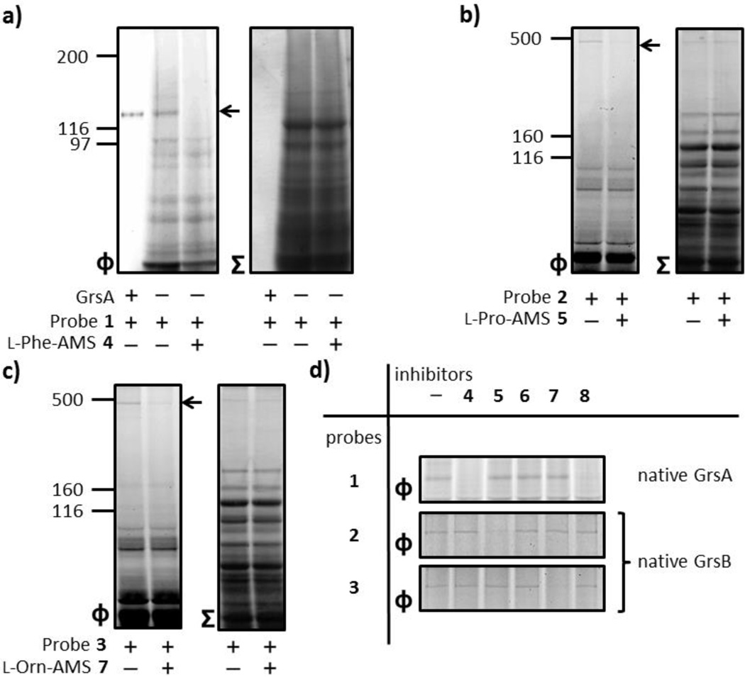
Proteomic applications of active site-directed proteomic probes 1–3 for A domains. (a) Labeling of endogenous GrsA in an A. migulanus ATCC 9999 cellular lysate by probe 1. The A. migulanus ATCC 9999 lysate (1.5 mg/mL) was treated with 1 µM 1 in either the absence or presence of 100 µM 4. (b) Labeling of endogenous GrsB in the A. migulanus DSM 5759 cellular lysate by probes 2 and 3. The A. migulanus DSM 5759 lysate (1.5 mg/mL) was individually treated with 100 µM 5 and 7 before the addition of individual members of 1 µM probes 2 and 3. (d) Individual labeling of A domains and profiling of A domain functions using a combination of probes 1–3 and inhibitors 4–8. In order to investigate GrsA labeling, the A. migulanus ATCC 9999 lysate (1.5 mg/mL) was preincubated with individual members of inhibitors 4–8 (100 µM) before the addition of 1 µM of probe 1. To evaluate the labeling of GrsB, the A. migulanus DSM 5759 lysate (1.5 mg/mL) was individually treated with 100 µM of inhibitors 4–8 before the addition of individual members of 1 µM probes 2 and 3. For each panel, Φ depicts the fluorescence observed with λex = 532 nm and λem = 580 nm, and Σ displays the total protein content by staining with Coomassie Blue. Full gels (Figure S4) and experimental procedures are provided in the SI.
Similar articles
-
Functional Diversity and Engineering of the Adenylation Domains in Nonribosomal Peptide Synthetases.Mar Drugs. 2024 Jul 29;22(8):349. doi: 10.3390/md22080349. Mar Drugs. 2024. PMID: 39195464 Free PMC article. Review.
-
Profiling Nonribosomal Peptide Synthetase Activities Using Chemical Proteomic Probes for Adenylation Domains.ACS Chem Biol. 2015 Sep 18;10(9):1989-97. doi: 10.1021/acschembio.5b00097. Epub 2015 Jun 17. ACS Chem Biol. 2015. PMID: 26038981
-
Accurate Detection of Adenylation Domain Functions in Nonribosomal Peptide Synthetases by an Enzyme-linked Immunosorbent Assay System Using Active Site-directed Probes for Adenylation Domains.ACS Chem Biol. 2015 Dec 18;10(12):2816-26. doi: 10.1021/acschembio.5b00595. Epub 2015 Oct 16. ACS Chem Biol. 2015. PMID: 26474351
-
A Multiple-Labeling Strategy for Nonribosomal Peptide Synthetases Using Active-Site-Directed Proteomic Probes for Adenylation Domains.Chembiochem. 2015 Dec;16(18):2590-4. doi: 10.1002/cbic.201500481. Epub 2015 Nov 6. Chembiochem. 2015. PMID: 26467472
-
Engineered Biosynthesis through the Adenylation Domains from Nonribosomal Peptide Synthetases.Curr Top Med Chem. 2023;23(20):1973-1984. doi: 10.2174/1568026623666230601142757. Curr Top Med Chem. 2023. PMID: 37264622 Review.
Cited by
-
Functional Diversity and Engineering of the Adenylation Domains in Nonribosomal Peptide Synthetases.Mar Drugs. 2024 Jul 29;22(8):349. doi: 10.3390/md22080349. Mar Drugs. 2024. PMID: 39195464 Free PMC article. Review.
-
Refining and expanding nonribosomal peptide synthetase function and mechanism.J Ind Microbiol Biotechnol. 2019 Mar;46(3-4):493-513. doi: 10.1007/s10295-018-02130-w. Epub 2019 Jan 23. J Ind Microbiol Biotechnol. 2019. PMID: 30673909 Free PMC article. Review.
-
Developing crosslinkers specific for epimerization domain in NRPS initiation modules to evaluate mechanism.RSC Chem Biol. 2022 Jan 27;3(3):312-319. doi: 10.1039/d2cb00005a. eCollection 2022 Mar 9. RSC Chem Biol. 2022. PMID: 35359491 Free PMC article.
-
Activity-based protein profiling of a surfactin-producing nonribosomal peptide synthetase in Bacillus subtilis.STAR Protoc. 2022 Jun 13;3(3):101462. doi: 10.1016/j.xpro.2022.101462. eCollection 2022 Sep 16. STAR Protoc. 2022. PMID: 35719724 Free PMC article.
-
Mining for Microbial Gems: Integrating Proteomics in the Postgenomic Natural Product Discovery Pipeline.Proteomics. 2018 Sep;18(18):e1700332. doi: 10.1002/pmic.201700332. Epub 2018 Jun 10. Proteomics. 2018. PMID: 29708658 Free PMC article. Review.
References
Publication types
MeSH terms
Substances
Grants and funding
LinkOut - more resources
Full Text Sources
Other Literature Sources
Molecular Biology Databases