. 2015 Feb 13;116(4):600-11.
doi: 10.1161/CIRCRESAHA.116.304899. Epub 2015 Jan 6.

Receptor-interacting protein kinase 3 contributes to abdominal aortic aneurysms via smooth muscle cell necrosis and inflammation

Affiliations

Receptor-interacting protein kinase 3 contributes to abdominal aortic aneurysms via smooth muscle cell necrosis and inflammation

Qiwei Wang et al. Circ Res. .

Abstract

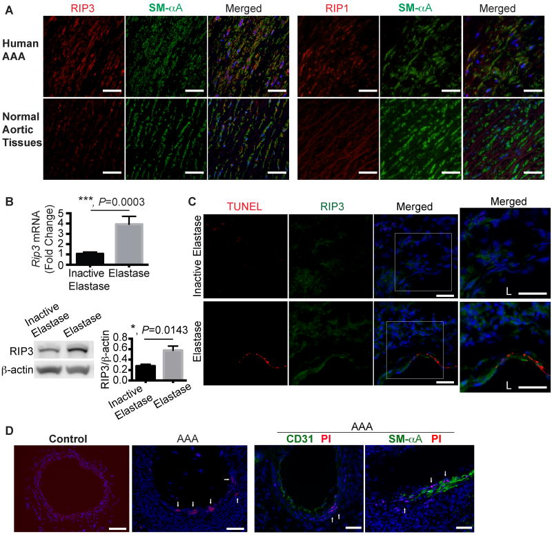
Rationale: Depletion of medial smooth muscle cell (SMC) is a major pathological characteristic of abdominal aortic aneurysm (AAA), although the mechanism by which these cells are eliminated remains incompletely understood. We reasoned that necroptosis, a recently described form of necrosis mediated by receptor-interacting protein kinase 3 (RIP3), may contribute to AAA pathology through the induction of SMC death and the significant production of inflammatory cytokines.

Objective: To test the hypothesis that RIP3-mediated necroptosis is actively involved in aneurysm pathogenesis.

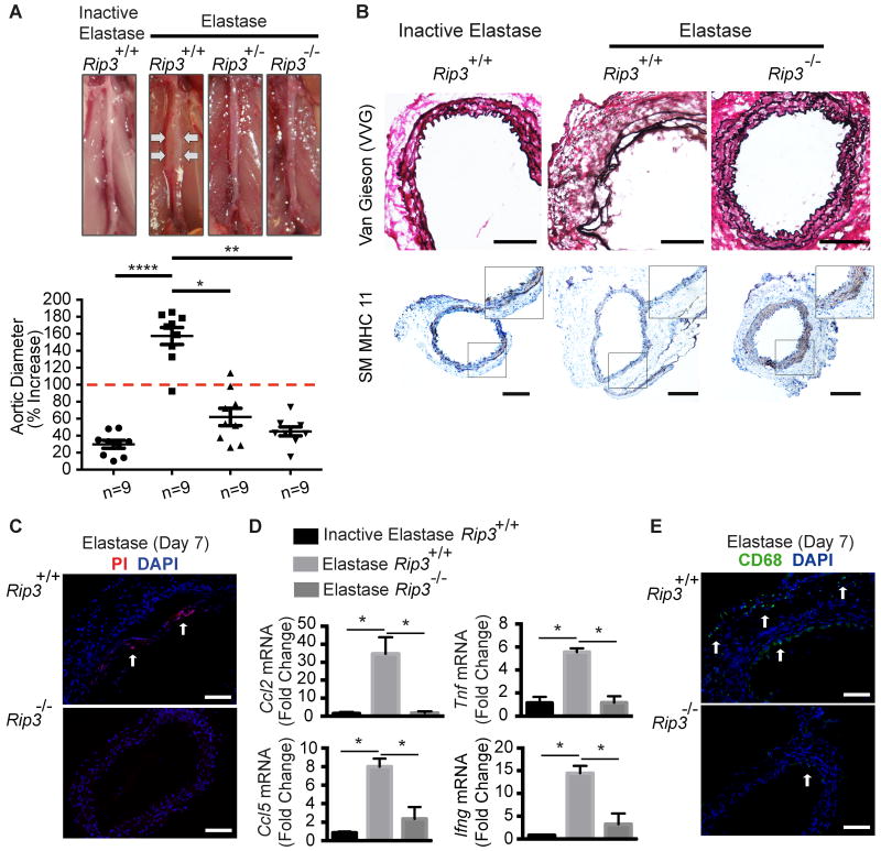
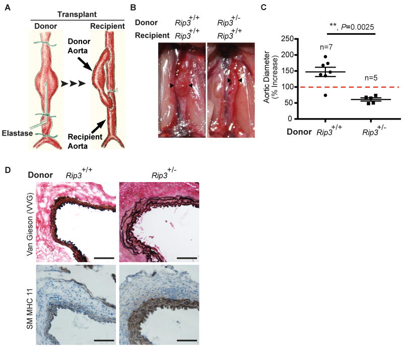
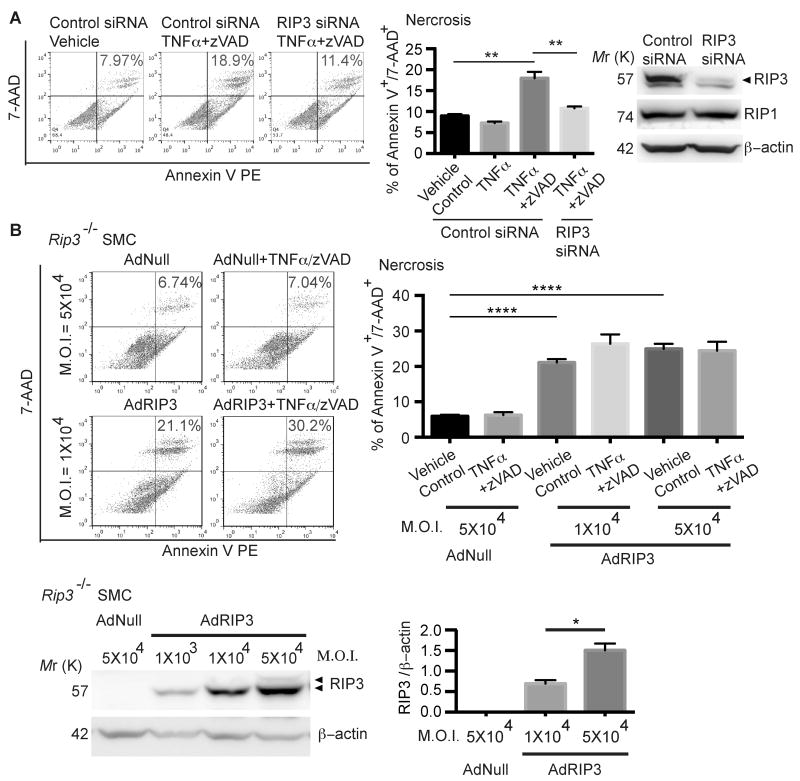
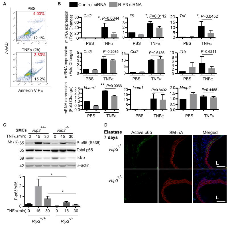
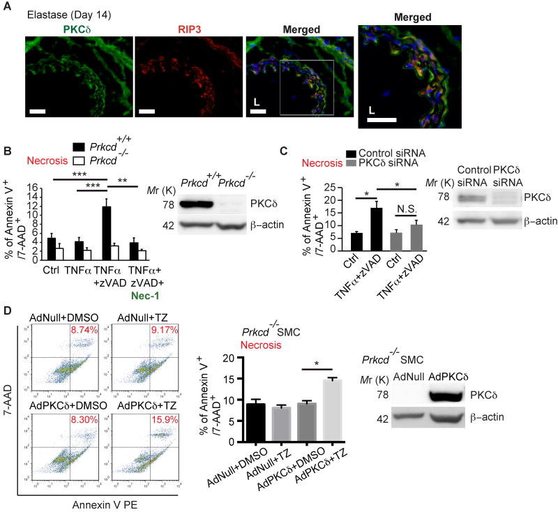
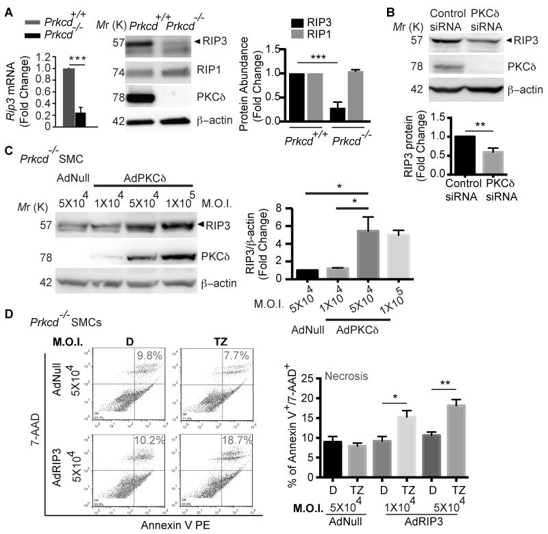
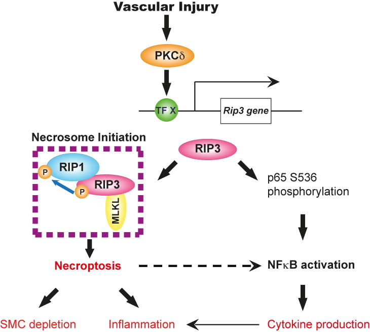
Methods and results: RIP3 and RIP1 levels were found to be elevated in human AAAs, most noticeably in SMCs. Elevations of RIP3 and SMC necrosis were also observed in the elastase-induced mouse model of AAAs. Deletion of one or both copies of Rip3 prevented AAA formation. By transplanting Rip3(+/-) aortae to Rip3(+/+) mice, we demonstrated that reduced Rip3 expression in arterial wall was the primary cause of aneurysm resistance. In vitro, adenoviral overexpression of RIP3 was sufficient to trigger SMC necroptosis. Protein kinase C-delta contributed to tumor necrosis factor-α-induced SMC necroptosis by regulating Rip3 expression. Furthermore, Rip3 deficiency impaired tumor necrosis factor-α-induced inflammatory gene expression in aortic SMCs, which was at least in part because of attenuation of p65 Ser536 phosphorylation. In vivo, the lack of RIP3 diminished activation of p65 in SMCs, implicating a necrosis independent function of RIP3 in aneurysms.

Conclusions: Enhanced RIP3 signaling in aneurysmal tissues contributes to AAA progression by causing SMC necroptosis, as well as stimulating vascular inflammation, and therefore may serve as a novel therapeutic target for AAA treatment.

Keywords: Rip3 protein, mouse; aortic aneurysm, abdominal; apoptosis; myocytes, smooth muscle; necrosis; nuclear factor kappa B; protein kinase C-delta.


Figures

Figure 1
Figure 1. Increased receptor interacting protein 3 (RIP3) expression and cell necrosis in abdominal aortic aneurysm tissues
(A) Representative photographs of human aneurysmal and normal aortic cross sections that were double stained for RIP3 (or RIP1, red) and smooth muscle α-Actin (SM-αA, green). n=4. Scale bars=50μm. (B) Real-time PCR (n=8) and Western blotting (n=4) analyses of Rip3 expression in C57BL/6 mouse arteries perfused with elastase or heat-inactivated elastase on Day 14 post-perfusion. Data are mean±SEM. (C) Representative photographs of aortic cross-sections harvested from C57BL/6 mice on Day 3 post-surgery. Sections were co-stained for RIP3 and TUNEL. Higher magnified views of highlighted regions were shown on the right. L indicates lumen. n=3. Scale bars=50μm. (D) Representative photographs of aortic cross sections of normal or elastase-perfused aortae (Day 7 post-perfusion). Propidium iodide (PI) was administered to mice via intraperitoneal injection 2 hours prior to sacrifice. Following PI staining, aortic sections were also stained for CD31 or SM-αA. n=3. Scale bars=100μm.
Figure 2
Figure 2. Rip3 deficiency protects mice from developing abdominal aortic aneurysm
(A) Mice of three genotypes were subjected to aneurysm induction by elastase perfusion. Animals were sacrificed 14 days after. Upper: Representative photos of abdominal aortae taken during sacrifice. Bottom: Aortic dilatation measured as the percentage increase of maximal external aortic diameter between pre-perfusion and on day 14 after perfusion. An AAA is defined as a percentage increase in aortic diameter greater than 100% (red dashed line). n=9. (B) Cross sections of harvested aortae were stained for Verhoeff-Van Gieson (VVG) in which elastin fibers are stained in dark purple or for smooth muscle myosin heavy chain 11 (SM MHC11). Inset photos are higher magnified views of highlighted regions. n=3. Scale bars=200μm. (C) Representative photos of aortic sections from mice that were injected with propidium iodide (PI) 2 hours prior to sacrifice on Day 7 post elastase perfusion. The necrotic cells (PI-positive) were indicated by white arrows. n=3. Scale bars=100μm. (D) Rip3+/+ and -/- mice were subjected to elastase perfusion and sacrificed 14 days after. Aortic levels of pro-inflammatory cytokines were analyzed by real-time PCR. n=4. (E) Representative photographs of immunostaining for a macrophage marker CD68 (green, arrows) in the elastase-perfused aortae. n=3. Scale bars=100μm. *P<0.05; **P<0.005; ****P<0.0001. All values represent mean±SEM.
Figure 3
Figure 3. Rip3 in the vascular wall is necessary for abdominal aortic aneurysm formation
(A) Graphic illustration of a mouse aortic transplant model. Prior to tissue transplantation, donor abdominal aorta was subjected to aneurysm induction via elastase perfusion for 5 minutes. Injured aortic segment was excised immediately following perfusion and anastomosed to a recipient abdominal aorta in an end-to-side fashion. (B) Representative photos taken 14 days after transplantation. Donor aortae are indicated by arrowheads. (C) Maximal external diameters of donor aortae were measured at sacrifice and prior to elastase perfusion. Aortic dilatation calculated as the percentage increase of maximal external aortic diameter between pre-perfusion and on day 14 after perfusion. An AAA is defined as a percentage increase in aortic diameter greater than 100% (red dashed line). Data represent mean±SEM. n=5∼7. (D) Cross sections of harvested aortae were stained for Verhoeff-Van Gieson (VVG) in which elastin fibers are stained in dark or for smooth muscle myosin heavy chain 11 (SM MHC11). n=3. Scale bars=50μm.
Figure 4
Figure 4. Aortic smooth muscle cells (SMCs) undergo RIP3-dependent necroptosis in vitro
(A) Aortic SMCs isolated from C57BL/6 mice were transfected with control siRNA or RIP3 siRNA. Cells were treated with DMSO (vehicle control), TNFα or TNFα plus zVAD for 24 hours. Cells were stained with PE Annexin V and 7-AAD and analyzed by flow cytometry. Necrotic cells were identified as PE Annexin V+/7-AAD+. The Western blot shown on the rightmost confirms the gene silence efficacy and specificity of RIP3 siRNA. Cell lysates were collected 36 hours post-transfection. **P<0.005. Data represent mean±SEM. n=3. (B) Rip3-/- aortic SMCs were infected with the empty adenoviral vector (AdNull) or the recombinant adenoviral vector expressing RIP3 (AdRIP3) at indicated multiplicity of infection (M.O.I.) units. After 24 hours, cells were treated with DMSO (vehicle control) or TNFα plus zVAD for 24 hours. Cell necrosis was determined by flow cytometry following staining with PE Annexin V and 7-AAD. Necrotic cells were identified as PE Annexin V+/7-AAD+. AdRIP3-mediated RIP3 expression in Rip3-/- SMCs was confirmed by Western blot. *P<0.05, ****P<0.0001. Data represent mean±SEM. n=3∼5.
Figure 5
Figure 5. RIP3 is involved in TNFα-induced cytokine expression in aortic SMCs
(A) Aortic SMCs isolated from C57BL/6 mice were treated with TNFα (10ng/ml) for 2 h. The absence of cell death induced by TNFα at this concentration was confirmed by flow cytometry. n=2. (B) Aortic SMCs were transfected with control or RIP3-specific siRNA. After 48 hours, cells were stimulated with 10ng/ml TNFα for 2 hours for cytokines and Mmp2 or 2ng/ml TNFα for 4 hours for Vcam1 and Icam1. mRNA levels were determined by Real-time PCR. Data are mean±SEM. n=4. (C) Aortic SMCs isolated form Rip3+/+ and Rip3-/- mice were treated with 10ng/mL TNFα for indicated time. Cell lysates were subjected to Western blot analysis with indicated antibodies. *P<0.05. Data represent mean±SEM. n=4. (D) Representative photographs of aortic cross-sections harvested from mice on Day 7 post-perfusion with elastase. Sections were co-stained for active p65 and SM-αA. L indicates lumen. n=3. Scale bars=50μm.
Figure 6
Figure 6. Prkcd -/- SMCs are resistant to necroptosis induction.
(A) Representative photographs of double immunostaining with PKCδ (green) and RIP3 (red) in elastase-perfused aortae harvested 14 days after surgery. Higher magnifications of highlighted regions are shown on the right. n=3. Scale bars=50μm. (B) Prkcd+/+ and Prkcd-/- SMCs were treated with DMSO (vehicle control), TNFα, TNFα plus zVAD with or without necrostatin-1 for 24 hours. Cell necrosis was determined by flow cytometric analysis following PE Annexin V and 7-AAD staining. Necrotic cells were identified as dual positive for both PE Annexin V and 7-AAD. Quantification of Necrosis (% of PE Annexin-V+/7-AAD+) is shown. The Western blot confirms the lack of PKCδ protein in Prkcd-/- SMCs. (C) Aortic SMCs were transfected with control siRNA or PKCδ-specific siRNA for 24 hours followed by treatment with DMSO (vehicle control) or TNFα plus zVAD for additional 24 hours. Cell necrosis was determined as described in (B). The efficacy of PKCδ knockdown was confirmed by Western blot. (D) Prkcd-/- aortic SMCs were infected with AdNull or AdPKCδ at M.O.I. of 5×10. Expression of PKCδ was analyzed by Western blot (rightmost panel). 36 hours post-infection, cells were treated with DMSO or TNFα plus zVAD (TZ). Cell necrosis was determined as described in (B). *P<0.05; **P<0.005; ***P<0.0005. Data represent mean±SEM. n=3 (B and D), n=5 (C).
Figure 7
Figure 7. PKCδ regulates necroptosis through controlling RIP3 expression
(A) Aortic SMCs isolated from Prkcd+/+ and Prkcd-/- mice were analyzed for Rip3 expression by real-time PCR and Western blot. ***P<0.0005. Data represent mean±SEM. n=3. (B) Aortic SMCs isolated from C57BL/6 mice were transfected with control siRNA or PKCδ siRNA. Cell lysates were collected 48 hours post-transfection and aliquots of 30μg protein were subjected for Western blot analysis of RIP3, PKCδ, and β-actin. Data represent mean±SEM. n=6. (C) Prkcd-/- aortic SMCs were infected with AdNull or AdPKCδ at indicated M.O.I. units. Cell lysates were collected 48 hours post-infection and aliquots of 30μg protein were analyzed by Western blot with indicated antibodies. Data represent mean±SEM. n=3. (D) Prkcd-/- aortic SMCs were infected with AdNull or AdRIP3. 48 hours post-infection, cells were treated with DMSO (D, vehicle control) or TNFα plus zVAD (TZ) for 24 hours. Cell necrosis was determined by flow cytometric analysis following PE Annexin V and 7-AAD staining. Necrotic cells were identified as PE Annexin V+/7-AAD+. Data represent mean±SEM. n=6 (D). *P<0.05; **P<0.005; ***P<0.0005.
Figure 8
Figure 8. Proposed roles of RIP3 in pathophysiology of abdominal aortic aneurysm
RIP3 is upregulated in the injured aneurysmal wall via a PKCδ-mediated mechanism (TF X: Transcription factor X). In the presence of elevated RIP3, SMCs respond to TNFα or other cytokines by undergoing necroptosis or NFκB-mediated chemokine/cytokine production. The precise determinants that switch RIP3 between necroptosis and NFκB in SMCs remain unclear, however, both processes may contribute to inflammation in aortic wall during AAA development and progression.

References

    1. Baxter BT, Terrin MC, Dalman RL. Medical management of small abdominal aortic aneurysms. Circulation. 2008;117:1883–1889. - PMC - PubMed
    1. Weintraub NL. Understanding abdominal aortic aneurysm. N Engl J Med. 2009;361:1114–1116. - PMC - PubMed
    1. Longo GM, Xiong W, Greiner TC, Zhao Y, Fiotti N, Baxter BT. Matrix metalloproteinases 2 and 9 work in concert to produce aortic aneurysms. The Journal of Clinical Investigation. 2002;110:625–632. - PMC - PubMed
    1. Pyo R, Lee JK, Shipley JM, Curci JA, Mao D, Ziporin SJ, Ennis TL, Shapiro SD, Senior RM, Thompson RW. Targeted gene disruption of matrix metalloproteinase-9 (gelatinase b) suppresses development of experimental abdominal aortic aneurysms. The Journal of Clinical Investigation. 2000;105:1641–1649. - PMC - PubMed
    1. Shimizu K, Mitchell RN, Libby P. Inflammation and cellular immune responses in abdominal aortic aneurysms. Arterioscler Thromb Vasc Biol. 2006;26:987–994. - PubMed

Publication types

MeSH terms