. 2015 Mar;35(3):512-20.
doi: 10.1038/jcbfm.2014.226. Epub 2015 Jan 7.

Simvastatin restored vascular reactivity, endothelial function and reduced string vessel pathology in a mouse model of cerebrovascular disease

Affiliations

Simvastatin restored vascular reactivity, endothelial function and reduced string vessel pathology in a mouse model of cerebrovascular disease

Xin-Kang Tong et al. J Cereb Blood Flow Metab. 2015 Mar.

Abstract

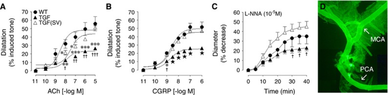
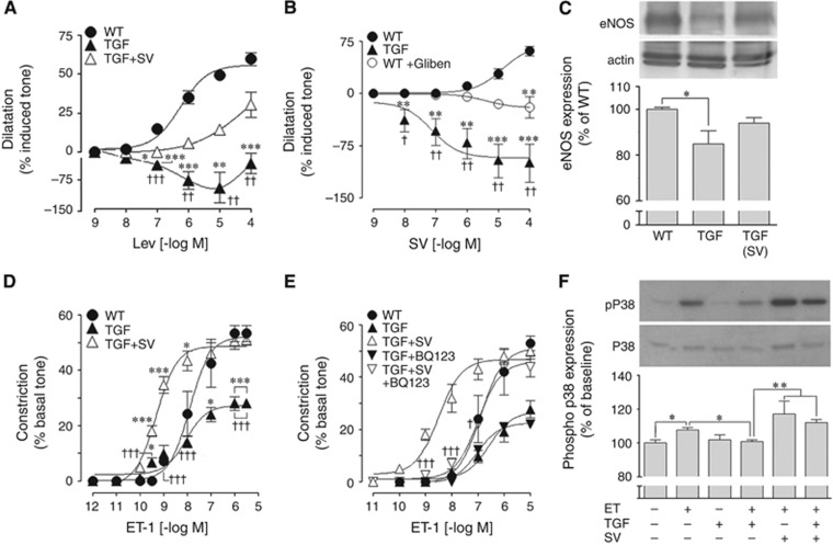
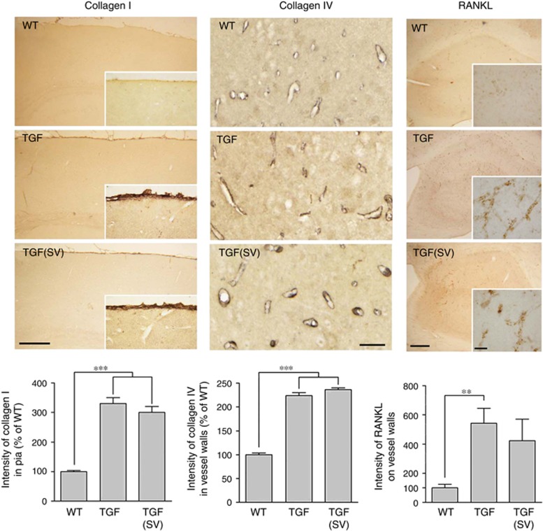
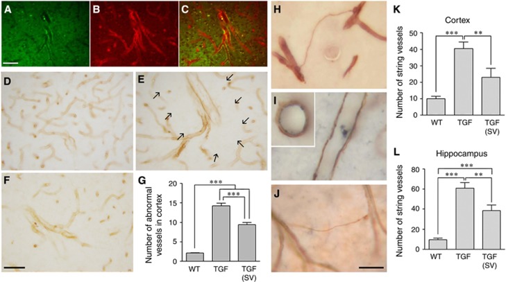
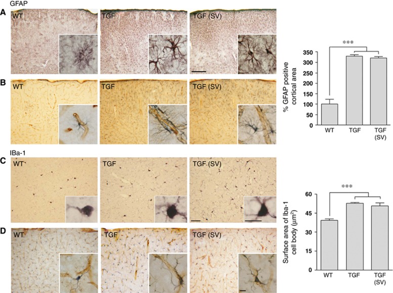
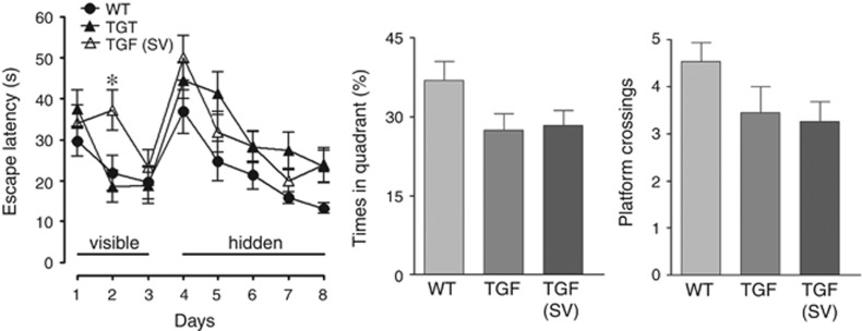
Cerebrovascular dysfunction seen in Alzheimer's disease (AD) and vascular dementia (VaD) is multifaceted and not limited to the amyloid-β (Aβ) pathology. It encompasses structural alterations in the vessel wall, degenerating capillaries (string vessels), vascular fibrosis and calcification, features recapitulated in transgenic mice that overexpress transforming growth factor-β1 (TGF mice). We recently found that simvastatin rescued Aβ-mediated cerebrovascular and cognitive deficits in a transgenic mouse model of AD. However, whether simvastatin can counteract Aβ-independent deficits remains unknown. Here, we evaluated the effects of simvastatin in aged TGF mice on cerebrovascular reactivity and structure, and on cognitive performance. Simvastatin restored baseline levels of nitric oxide (NO), NO-, and KATP channel-mediated dilations and endothelin-1-induced contractions. Simvastatin significantly reduced vasculopathy with arteriogenic remodeling and string vessel pathology in TGF mice. In contrast, simvastatin did not lessen gliosis, and the cerebrovascular levels of pro-fibrotic proteins and calcification markers remained elevated after treatment. The TGF mice displayed subtle cognitive decline that was not affected by simvastatin. Our results show potent benefits of simvastatin on endothelial- and smooth muscle cell-mediated vasomotor responses, endothelial NO synthesis and in preserving capillary integrity. We conclude that simvastatin could be indicated in the treatment of cerebrovascular dysfunction associated with VaD and AD.


Figures

Figure 1
Figure 1
Simvastatin (SV) improved cerebrovascular reactivity in transforming growth factor-β1 (TGF) mice. The impaired dilatory responses of isolated arteries to acetylcholine (ACh) (A) and calcitonin gene-related peptide (CGRP) (B) in TGF mice (▴, n=5) relative to wild-type controls (WT, ●, n=3) were normalized in SV-treated TGF (△, n=5) mice. Similarly, the basal nitric oxide (NO) synthesis, measured through NO synthase (NOS) inhibition with Nω-nitro-L-arginine (L-NNA) (C, 10−5 mol/L) was fully restored by SV treatment (△). TGF mice did not display perivascular amyloid deposits as seen with Thioflavin-S staining, the brighter segments correspond to small pieces of the attached pial membrane (D). MCA, middle cerebral artery; PCA, posterior cerebral artery. *P<0.05, **P<0.01, ***P<0.001 compared with WT; P<0.05, ††P<0.01, †††P<0.001 compared with SV-treated TGF mice; P<0.05 compared with WT and SV-treated TGF mice.
Figure 2
Figure 2
Simvastatin (SV) improved KATP channel function, endothelial nitric oxide synthase (eNOS) protein levels, and potentiated endothelin-1 (ET-1) type A (ETA) receptor function in transforming growth factor-β1 (TGF) mice through activation of p38 MAP kinase. (A) KATP channels were impaired in TGF mice (▴, n=6) as shown with the channel opener levcromakalim (Lev). SV restored KATP channel function in treated TGF mice (△, n=4) to levels not significantly different from wild-type (WT) controls (●, n=4). (B) Impaired KATP channel function in TGF mice (▴, n=4) when activated by SV was mimicked in WT arteries incubated with the KATP channel blocker glibenclamide (Gliben, ○, n=6), vessels showing contraction to SV instead of dilation. (C) Western blot analysis of eNOS protein showed significantly reduced levels in vessels from TGF mice compared with WT controls, and SV increased these to levels not significantly different from WT (n=4 to 7 mice/group). (D) The impaired contractile response to ET-1 in TGF mice (▴, n=5) compared with WT controls (●, n=3) was potentiated beyond control levels in SV-treated TGF mice (△, n=4). This SV potentiating effect was completely blocked by the selective ETA receptor antagonist BQ123 (E), which had no effect in vessels from non-treated TGF mice. (F) The ET-1-induced phosphorylation of p38 MAP kinase in cerebral smooth muscle cell cultures was inhibited by TGF-β1, an effect prevented by preincubation with SV (n=3 to 6). *P<0.05; **P<0.01; ***P<0.001 compared with WT; P<0.05; ††P<0.01; †††P<0.001 compared with SV-treated TGF mice, or WT+glibemclamide.
Figure 3
Figure 3
Simvastatin (SV) failed to normalize vascular fibrosis and slightly reduced vascular calcification. The intensity of immunostaining for collagen I (n=3 mice/group) in the pial membrane and of collagen IV (n=4 mice/group) in the walls of cortical microvessels in 5m-thick paraffin sections was increased in transforming growth factor-β1 (TGF) mice compared with wild-type (WT) controls, and SV exerted no reducing effect at this level. In 25-μm-thick hippocampal sections, the intensity of receptor activator of nuclear factor κB ligand (RANKL) immunopositive vessels was significantly increased in TGF mice (n=5) compared with WT controls (n=4), and SV (n=6) slightly reduced this intensity to levels not significantly different from WT controls. Bars: left panel=250 μm; middle panel=50 μm; and right panel=200 μm (inset=20 μm). **P<0.01; ***P<0.001.
Figure 4
Figure 4
Simvastatin (SV) reduced arteriogenic remodeling and string vessel pathology in transforming growth factor-β1 (TGF) mice. (A to C) Double immunofluorescence for the endothelial cell marker CD31 (A, green) and smooth muscle actin (B, red) showed the presence of abnormally large intracortical arterioles (C, merge). These large vessels immunopositive for collagen IV were rarely seen in the cortex of WT controls (D), but they were numerous in TGF mice (E and F). In most cases, these large vessels had few ramifications and were surrounded by an area devoid of capillaries (arrows in E, see also Figure 5). SV treatment slightly but significantly reduced this pathology (G) (n=3 mice/group). (H) String vessel pathology was revealed by collagen IV immunostaining in TGF mice. In contrast to WT capillaries that show endothelial CD31 immunostaining (I, blue) inside the collagen IV immunostained basement membrane (I, brown), the thin walls of string vessels only stained for collagen IV (H and J). SV treatment significantly reduced the number of string vessels in cortex and hippocampus of TGF mice (K and L) (n=7 to 9 mice/group). **P<0.01; ***P<0.001. Bars: (A to C)=50 μm, (D to F)=30 μm.
Figure 5
Figure 5
Simvastatin (SV) slightly increased the number of capillaries around large intracortical arterioles in transforming growth factor-β1 (TGF) mice. Histochemical NADPH diaphorase staining of cortical blood vessels in TGF (n=8) and SV-treated (n=9) TGF mice showed that SV slightly but significantly increased the number of small vessels (open arrows) localized in the immediate vicinity (dotted lines) of large intracortical arterioles. However, SV did not improve the limited branching (black arrows) of these large vessels. P<0.05 by Student's t-test. Bar=30 μm.
Figure 6
Figure 6
Simvastatin (SV) did not suppress astrogliosis and microgliosis in the cortex of transforming growth factor-β1 (TGF) mice. Glial fibrillary acidic protein (GFAP)-immunostained astrocytes in the cerebral cortex (A) and double immunostaining for intracortical microvessels and astrocytes (B, collagen IV, (brown: vessels) and GFAP (gray: astrocytes)) showed an activated astroglial phenotype with larger cell bodies and higher number of processes. The surface area occupied by GFAP-positive astrocytes was increased in TGF mice compared with WT controls, and SV has no effect at this level (top right panel) (n=4 to 6 mice/group). Similarly, Iba-1 immunopositive microglial cells displayed a reactive phenotype with increased processes (C), some of which associated with intracortical microvessels (D). SV did not reduce this phenotype (n=4 mice/group). Bars: top panel=200 μm and bottom=50 μm. Inserts, Bar=10 μm. ***P<0.001.
Figure 7
Figure 7
Aged transforming growth factor-β1 (TGF) mice exhibited no significant deficit in the Morris water maze, and simvastatin (SV) had not effect on cognitive performance. TGF mice and SV-treated TGF mice did not significantly differ from wild-type (WT) controls in the time they needed to find the visible or hidden platform (left panel), despite the longer escape latency needed by SV-treated TGF mice on day 2 of the visible platform training. Similarly, TGF and SV-treated TGF mice were slightly but not significantly impaired in the probe trial (n=15 mice/group).

References

    1. Iadecola C. The pathobiology of vascular dementia. Neuron. 2013;80:844–866. - PMC - PubMed
    1. Wiesmann M, Kiliaan AJ, Claassen JA. Vascular aspects of cognitive impairment and dementia. J Cereb Blood Flow Metab. 2013;33:1696–1706. - PMC - PubMed
    1. Iadecola C, Zhang F, Niwa K, Eckman C, Turner SK, Fischer E, et al. SOD1 rescues cerebral endothelial dysfunction in mice overexpressing amyloid precursor protein. Nat Neurosci. 1999;2:157–161. - PubMed
    1. Tong XK, Lecrux C, Hamel E. Age-dependent rescue by simvastatin of Alzheimer's disease cerebrovascular and memory deficits. J Neurosci. 2012;32:4705–4715. - PMC - PubMed
    1. Brown WR, Thore CR. Review: cerebral microvascular pathology in ageing and neurodegeneration. Neuropathol Appl Neurobiol. 2011;37:56–74. - PMC - PubMed

Publication types

Substances

LinkOut - more resources