. 2015 Feb 18;27(6):064113.
doi: 10.1088/0953-8984/27/6/064113. Epub 2015 Jan 7.

Forced unraveling of chromatin fibers with nonuniform linker DNA lengths

Affiliations

Forced unraveling of chromatin fibers with nonuniform linker DNA lengths

Gungor Ozer et al. J Phys Condens Matter. .

Abstract

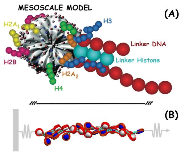
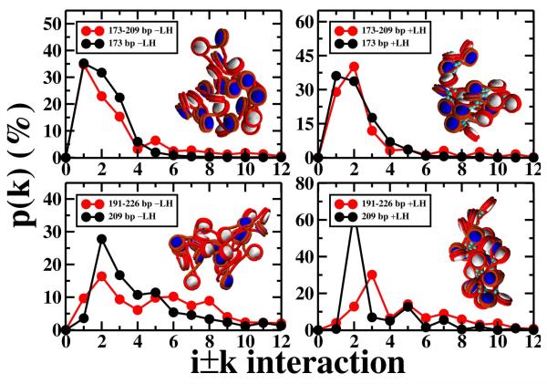
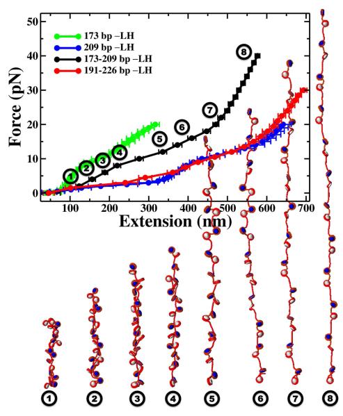
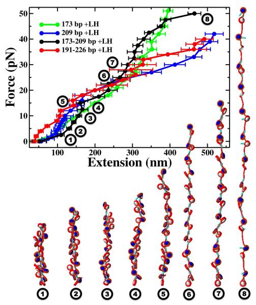
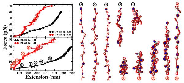
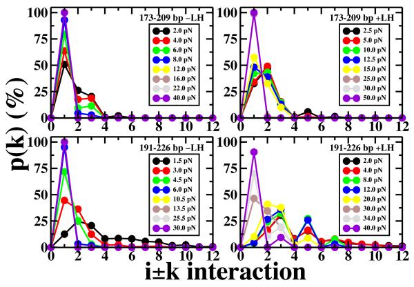
The chromatin fiber undergoes significant structural changes during the cell's life cycle to modulate DNA accessibility. Detailed mechanisms of such structural transformations of chromatin fibers as affected by various internal and external conditions such as the ionic conditions of the medium, the linker DNA length, and the presence of linker histones, constitute an open challenge. Here we utilize Monte Carlo (MC) simulations of a coarse grained model of chromatin with nonuniform linker DNA lengths as found in vivo to help explain some aspects of this challenge. We investigate the unfolding mechanisms of chromatin fibers with alternating linker lengths of 26-62 bp and 44-79 bp using a series of end-to-end stretching trajectories with and without linker histones and compare results to uniform-linker-length fibers. We find that linker histones increase overall resistance of nonuniform fibers and lead to fiber unfolding with superbeads-on-a-string cluster transitions. Chromatin fibers with nonuniform linker DNA lengths display a more complex, multi-step yet smoother process of unfolding compared to their uniform counterparts, likely due to the existence of a more continuous range of nucleosome-nucleosome interactions. This finding echoes the theme that some heterogeneity in fiber component is biologically advantageous.


Figures

Figure 1
Figure 1
Chromatin model and setup. A) The basic building block of our mesoscale chromatin model is displayed on a single nucleosome with linker histone and alternating 44-79 bp linker DNA (corresponding to 4-8 beads in our coarse-grained resolution). The nucleosome core plus the wrapping DNA is modeled as uniformly distributed charges. Core histone tails, linker histones and linker DNAs are modeled explicitly as labeled. B) The pulling procedure is illustrated on the same fiber at the coarse-grained resolution.
Figure 2
Figure 2
Probability distributions of the i ± k nucleosome-nucleosome interactions for equilibrium conformation. Top panels represent data for alternating 173-209 bp and uniform 173 bp fibers; bottom panels correspond to alternating 191-226 bp NRL and uniform 209 bp fibers, respectively. The nonuniform 173-209 bp fiber is a “bent-ladder” with dominant ii ± 1, 2, 3 internucleosome interactions. The 191-226 fiber adopts a polymorphic structure with ii ± 2, 3, 5 contacts, different than its uniform counterpart, 209 bp fiber. The left and right panels correspond to models without linker histones and with linker histones, respectively.
Figure 3
Figure 3
Stretching response of chromatin fibers with alternating versus uniform linker DNA length in the absence of linker histones (−LH). Black and red represent the force-extension data for alternating 173-209 bp and 191-226 bp NRL fibers; green and blue represent the same data for the uniform 173 bp and 209 bp fibers, respectively. Error bars denote standard deviations from the mean end-to-end separation of each force-specific ensemble. The snapshots represent the minimum energy configuration selected from the equilibrium ensemble of the chromatin fiber stretched using the labeled (circled numbers) forces. The forces corresponding to snapshots 1–8 are 2, 4, 6, 8, 12, 16, 22, and 40 pN, respectively.
Figure 4
Figure 4
Stretching response of chromatin fibers with alternating versus uniform linker DNA length in the presence of linker histones (+LH). Black and red represent the force-extension data for alternating 173-209 bp and 191-226 bp NRL fibers; green and blue represent the same data for the uniform 173 bp and 209 bp fibers, respectively. Error bars denote standard deviations from the mean end-to-end separation of each force-specific ensemble. The snapshots represent the minimum energy configuration selected from the equilibrium ensemble of the chromatin fiber stretched using the labeled (circled numbers) forces. The forces corresponding to snapshots 1–8 are 2.5, 5, 10, 12.5, 15, 25, 30, and 50 pN, respectively.
Figure 5
Figure 5
Stretching response of chromatin fibers with alternating linker DNA lengths in the presence (red) and absence (black) of linker histones. Top and bottom panels represent data for 173-209 bp and 191-226 bp NRL fibers, respectively. Error bars denote standard deviations from the mean end-to-end separation of each force-specific ensemble. The snapshots (in black without LH and red with LH) correspond to minimum energy configuration selected from the equilibrium ensemble of the chromatin fiber with alternating 44-79 bp linker DNA length (or 191-226 bp in NRL units) stretched using the labeled (circled numbers) forces. The forces correspond to snapshots 1–5 are 1.5, 3, 4.5, 6, and 10.5 pN for the −LH case; 2, 4, 8, 20, and 30 pN for the +LH case (red), respectively.
Figure 6
Figure 6
Probability distributions of the i ± k nucleosome-nucleosome interactions for equilibrium fibers at different applied forces. Top panels represent data for the alternating 173-209 bp fiber, bottom panels correspond to the alternating 191-226 bp NRL fiber. The left and right panels correspond to models without histones and with linker histones, respectively.

Similar articles

Cited by

References

    1. Finch J, Klug A. Proc. Natl. Acad. Sci. U.S.A. 2011;73(6):1897. - PMC - PubMed
    1. Maeshima K, Hihara S, Eltsov M. Curr. Opin. Cell. Biol. 2010;22:291. - PubMed
    1. Eltsov M, MacLellan KM, Maeshima K, Frangakis AS, Dubochet J. Proc. Natl. Acad. Sci. U.S.A. 2008;105(50):19732. - PMC - PubMed
    1. McDowall A, Smith J, Dubochet J. EMBO J. 1986;5(6):1395. - PMC - PubMed
    1. Woodcock C, Frado L, Rattner J. J. Cell. Biol. 1984;99(1 Pt 1):42. - PMC - PubMed

Publication types

LinkOut - more resources