. 2015 Mar 5;125(10):1566-77.
doi: 10.1182/blood-2014-11-610816. Epub 2015 Jan 6.

Hemophagocytic lymphohistiocytosis caused by dominant-negative mutations in STXBP2 that inhibit SNARE-mediated membrane fusion

Affiliations

Hemophagocytic lymphohistiocytosis caused by dominant-negative mutations in STXBP2 that inhibit SNARE-mediated membrane fusion

Waldo A Spessott et al. Blood. .

Abstract

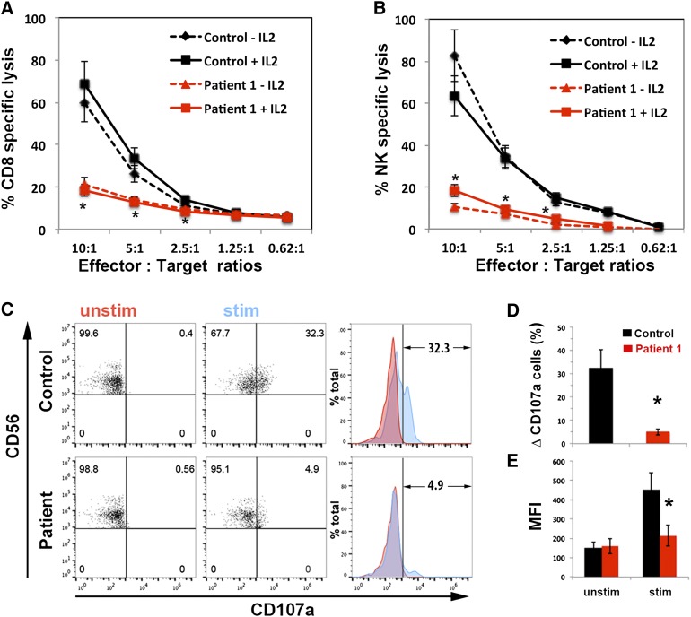
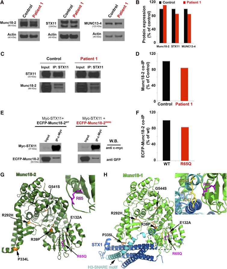
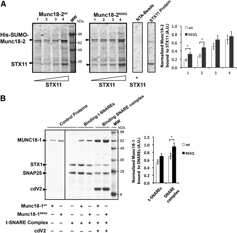
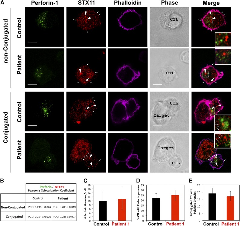
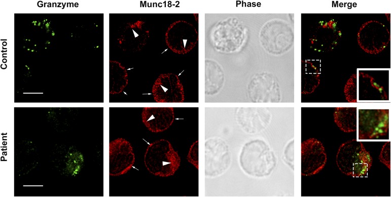
Familial hemophagocytic lymphohistiocytosis (F-HLH) and Griscelli syndrome type 2 (GS) are life-threatening immunodeficiencies characterized by impaired cytotoxic T lymphocyte (CTL) and natural killer (NK) cell lytic activity. In the majority of cases, these disorders are caused by biallelic inactivating germline mutations in genes such as RAB27A (GS) and PRF1, UNC13D, STX11, and STXBP2 (F-HLH). Although monoallelic (ie, heterozygous) mutations have been identified in certain patients, the clinical significance and molecular mechanisms by which these mutations influence CTL and NK cell function remain poorly understood. Here, we characterize 2 novel monoallelic hemophagocytic lymphohistiocytosis (HLH)-associated mutations affecting codon 65 of STXPB2, the gene encoding Munc18-2, a member of the SEC/MUNC18 family. Unlike previously described Munc18-2 mutants, Munc18-2(R65Q) and Munc18-2(R65W) retain the ability to interact with and stabilize syntaxin 11. However, presence of Munc18-2(R65Q/W) in patient-derived lymphocytes and forced expression in control CTLs and NK cells diminishes degranulation and cytotoxic activity. Mechanistic studies reveal that mutations affecting R65 hinder membrane fusion in vitro by arresting the late steps of soluble N-ethylmaleimide-sensitive factor attachment protein receptor (SNARE)-complex assembly. Collectively, these results reveal a direct role for SEC/MUNC18 proteins in promoting SNARE-complex assembly in vivo and suggest that STXBP2 R65 mutations operate in a novel dominant-negative fashion to impair lytic granule fusion and contribute to HLH.


Figures

Figure 1
Figure 1
CTLs and NK cells expressing the STXBP2R65Q mutation exhibit impaired functions. (A) Cytotoxicity assay to measure CTL-mediated cell killing. PBMCs from control (black) and P1 cells (red) were cultured with CD3/CD28 beads for 5 days and then incubated for 48 hours with (solid line) or without (dashed line) 200 U/mL of IL-2. Equivalent numbers of CD8+ T-cells (Effector) were purified from control and P1 PBMCs and then incubated with anti-CD3 antibody in the presence or absence of P815 target cells (Target) at the indicated cell ratios. The killing assay was run for 4 hours at 37°C, and the amount of lactate dehydrogenase released into the supernatant was quantified using a CytoTox 96 assay. (B) NK cytotoxicity was assessed using control and P1 PBMCs incubated with K562 target cells, as described in panel A. Target-cell lysis was normalized to the number of NK cells in the PBMCs to obtain the percent NK-specific lysis. (C) CD107a assay to measure degranulation. PBMCs from control and P1 were incubated in the presence (stim; blue) or absence (unstim; red) of K562 cells for 4 hours at 37°C. Cells were stained using anti-CD107a-PE, anti-CD56-APC, anti-CD8-FITC, and anti-CD3-PerCP antibodies and analyzed by flow cytometry. CD3CD8CD56+ cells were gated and analyzed for the appearance of CD107a on the surface after incubation with target cells. Plots are representative of 3 independent experiments. (D) Graph showing the percentage of cells that increased CD107a staining on stimulation. The term “δ CD107a” reflects the difference between the percentage of NK cells expressing CD107a after K562 stimulation and the percentage of cells expressing surface CD107a after incubation with medium. (E) Mean fluorescence intensity (MFI) values in the CD107a-PE channel of unstimulated (unstim) vs stimulated (stim) cells. Results are the mean ± SD of 3 independent measurements for P1 and 10 different normal controls. *P < .01.
Figure 2
Figure 2
The STXBP2 mutation does not influence protein expression or the Munc18-2/STX11 interaction. (A) Western blots showing the expression levels of Munc18-2, STX11, and MUNC13-4 in lysates prepared using PBMCs activated with CD3/28 beads from control (black) or P1 (red). Actin staining of the same membranes was used to assess for equivalent protein loading. (B) Bands in the western blot that corresponded to Munc18-2, STX11, and MUNC13-4 were quantified by densitometry and normalized to the intensity of actin in the same lane. Densitometry results are expressed as the percentage of those obtained using control samples, which were set as 100%. (C) Coimmunoprecipitation experiments using lysates generated from control or P1 PBMCs. Endogenous STX11 was immunoprecipitated (IP) using an anti-STX11 antibody, and the amount of Munc18-2 that coimmunoprecipitated was quantified by western blot analysis. (D) Bands in the western blot that corresponded to the fraction of Munc18-2 that coimmunoprecipitated (co-IP) with STX11 were quantified by densitometry and normalized to the amount of STX11 immunoprecipitated in the same lane. Densitometry results were expressed as the percentage of those obtained in control samples, which were set as 100%. (E) Coimmunoprecipitation experiments using HeLa cells transiently transfected with Myc-STX11 and ECFP-Munc18-2WT (black) or ECFP-Munc18-2R65Q (red). STX11 was immunoprecipitated (IP) using an anti-Myc antibody and the amount of Munc18-2 that coassociated was quantified by western blot analysis using an anti-GFP antibody. (F) Densitometry analysis was performed as described in panel C. In panels A-F, results are representative of 2 independent experiments. (G) Crystal structure of Munc18-2 (Protein Data Bank entry 4CCA) with the R65 residue highlighted in magenta. Previously described HLH-associated Munc18-2 missense mutants are highlighted in orange. (H) The crystal structure of Munc18-1 (green) in complex with STX1 (Habc domain: blue, H3 domain: cyan; Protein Data Bank entry 3C98) reveals that the C-terminal half of the STX1 H3 domain inserts deeply into the Munc18-1 central cavity. The R65 residue (highlighted in magenta) is in proximity to residues D242 and Y243 (yellow) within the STX1 H3 domain but it does not make direct contact with these residues. Munc18-1 residues corresponding to described missense mutations in Munc18-2 are highlighted in orange. The insets in G and H represent a higher magnification of the structure, rotated 90° clockwise in the x-axis.
Figure 3
Figure 3
The R65Q mutation does not affect binding of Munc18 proteins to syntaxins, t-SNAREs, or SNARE complexes. (A) Pull-down experiments in which equivalent amounts of recombinant His-SUMO-Munc18-2WT or Munc18-2R65Q were bound to nickel-nitrilotriacetic acid beads (NTA-Beads) and increasing concentrations of recombinant STX11 were added to the beads. Bound fractions were analyzed by sodium dodecyl sulfate–polyacrylamide gel electrophoresis and coomassie blue staining. Plot shows the amount of STX11 bound to the beads normalized by the amount of Munc18-2WT or R65Q present on the beads. (B) Pull-down experiments in which recombinant His-SNAP25/STX1 (t-SNAREs) or His-Vamp2/STX1/SNAP25 (SNARE complex) were immobilized on NTA-Beads and incubated in the presence of equivalent amounts of either untagged Munc18-1WT or Munc18-1R65Q. Bound fractions were analyzed by sodium dodecyl sulfate–polyacrylamide gel electrophoresis and coomassie blue staining. Plot shows the amount of Munc18-1WT or R65Q bound to t-SNAREs or SNARE complexes normalized by the amount of STX1 present on the beads. Gels and plots are representative of 2 independent experiments. *P < .05. A.U., arbitrary units; cdV2, soluble domain of Vamp2.
Figure 4
Figure 4
The R65Q mutation does not affect the subcellular localization of STX11 or the number and polarization of perforin-containing granules. (A) CTLs from a control individual or P1 were incubated in the presence or absence of anti-CD3-coated P815 cells at a 1:1 ratio for 15 minutes at 37°C on polylysine-coated coverslips. Cells were fixed, permeabilized, and stained using mouse anti-perforin-1 antibody, rabbit anti-STX11 antibody and Alexa 633-Phalloidin. Larger arrowheads show the intracellular vesicular pool of STX11. Smaller arrows show the fraction of STX11 localizing at the plasma membrane. Bars represent 5 μm. Insets display a zoomed-in view of the selected area. (B) Pearson’s colocalization coeficient (PCC) between perforin 1 and STX11 in the set of images shown in panel A. Values represent the mean ± standard deviation (SD); n = 15 cells. Quantification of the number of perforin-containing granules per cell (C) and the number of cells containing perforin granules (D) was performed using stimulated emission depletion images (as shown in panel A; nonconjugated). Data represent the mean ± SD; n = 25 cells for panel C and n = 100 cells for panel D. (E) Quantification of the number of CTLs making contact with target cells that display polarized granules. Stimulated emission depletion images (as shown in panel A; conjugated) were used to determine the percentage of CTLs in contact with target cells that exhibited polarized perforin-containing granules at the immunologic synapse among the total number of CTLs containing perforin granules. Data represent the mean ± SD; n = 50 cells.
Figure 5
Figure 5
The R65Q mutation does not affect the subcellular localization of Munc18-2. CTLs from a control or P1 were incubated for 15 minutes at 37°C on polylysine-coated coverslips. Cells were fixed, permeabilized, and stained using mouse anti-granzyme-A or rabbit anti-Munc18-2 antibody. Large arrowheads show the intracellular pool of Munc18-2. Smaller arrows show the fraction of Munc18-2 localizing at the plasma membrane. Insets display a zoomed-in view of the selected area. Bars represent 5 μm.
Figure 6
Figure 6
The R65Q mutation inhibits membrane fusion in vitro. (A) Schematic depicting the liposome fusion reaction. (B) T- and labeled v-liposomes were preincubated on ice for 2 hours (hs) with control buffer, 2.5 μM Munc18-1WT, 2.5 μM Munc18-1R65Q, or 2.5 μM cdV2. Subsequently, the temperature was increased to 37°C, and the fluorescent signal was read every 2 minutes (min) for a total of 120 minutes. (C) Dose-dependent inhibition of fusion by Munc18-1R65Q. The liposome fusion reaction was performed as in panel B but with increasing concentrations of Munc18-1R65Q. (D) Dose-dependent activation by Munc18-1WT. The liposome fusion reaction was performed as in panel B but with increasing concentrations of Munc18-1WT. (E) Competition experiments using Munc18-1WT and Munc18-1R65Q. T- and v-liposomes were preincubated on ice for 2 hours in the presence of 2.5 μM Munc18-1WT and control buffer or increasing concentrations of Munc18-1R65Q (1.2-5.0 μM). Subsequently, the temperature was increased to 37°C, and the fluorescent signal was read every 2 minutes for a total of 120 minutes. (F) Competition experiments using Munc18-1R65Q and Munc18-1WT. T- and v-liposomes were preincubated on ice for 1 hour in the absence (control) or presence of 2.5 μM Munc18-1R65Q. Munc18-1R65Q-treated liposomes were incubated on ice for 2 hours with increasing concentrations of Munc18-1WT (2.5-10.0 μM). The temperature was increased to 37°C, and fusion was monitored as described above. (G) T-liposomes were incubated in the absence (control) or presence of Munc18-1WT, Munc18-1R65Q, or cdV2 for 2 hours on ice, followed by the addition of v-liposomes. (H) T- and v-liposomes were preincubated for 2 hours on ice to allow the assembly of SNARE complexes, followed by the addition of control buffer, Munc18-1WT, or Munc18-1R65Q. Subsequently, the temperature was raised to 37°C, and lipid mixing was measured. (I) T- and v-liposomes were preincubated 2 hours on ice, and then cdV2 was added to the reaction and incubated for 1 hour on ice. After that, control buffer, Munc18-1WT, or Munc18-1R65Q was added, and the temperature was increased to 37°C to induce lipid mixing. In panels B-I, the blue curve shows the effect when the strong inhibitor cdV2 was added from the beginning of the fusion reaction. Graphs in the insets of panels B-I show the fold activation of at least 3 independent experiments using different liposome preparations. The fold activation was calculated as a ratio of the difference of the initial rate of membrane fusion at 60 minutes in the presence of Munc18-1 minus the initial rate of fusion in the absence of Munc18-1 divided by the initial rate of fusion in the absence of Munc18-1. *P < .05 compared to control level.
Figure 7
Figure 7
Expression of Munc18-2R65Q in control CTLs reduces cytolytic activity. (A) CD8+ T cells from normal control donors were transduced with lentiviral particles encoding for ECFP-Munc18-2R65Q, ECFP-Munc18-2WT, or ECFP alone. Seven days posttransduction, the cytotoxic activity against anti-CD3-coated P815 cells was tested at the indicated effector to target-cell ratios. (B) Expression levels of ECFP-Munc18-2R65Q and ECFP-Munc18-2wt were compared to endogenous Munc18-2 by western blot analysis using anti-Munc18-2 antibody. (C) CD107a degranulation assay for ECFP-expressing cells. ECFP+ cells were purified by cell sorting and were incubated in the presence (stim; blue) or absence (unstim; red) of CD3-coated P815 cells for 4 hours at 37°C. Cells were stained using anti-CD107a-PE, anti-CD56-APC, anti-CD8-FITC, and anti-CD3-PerCP antibodies and analyzed by flow cytometry. CD3+CD8+CD56 cells were gated and analyzed for the appearance of CD107a on the cell surface following incubation with target cells. Plots are representative of 2 independent experiments. (D) Graph showing the percentage of cells that increased CD107a staining after stimulation and the mean fluorescence intensity (MFI) values in the CD107a-PE channel of unstimulated (u) and stimulated (s) cells. Results are the mean ± standard deviation of 2 independent measurements. *P < .01. (E-F) Schematic representation depicting the mode of action of Munc18-2 during lytic granule secretion in a normal control (E) or patient cell carrying the STXBP2R65Q mutation (F). Step 1: soluble Munc18-2WT and Munc18-2R65Q can bind monomeric STX11 on the acceptor membrane (probably the plasma membrane). Step 2: lytic granules approach the plasma membrane on cell activation. Step 3: both Munc18-2WT and Munc18-2R65Q facilitate SNARE-complex assembly between specific v-SNAREs (such as Vamp8) and target-SNAREs (such as STX11 and SNAP23). Step 4: Munc18-2R65Q, but not Munc18-2WT, may arrest some SNARE complexes in a partially zippered state and thus uncouple their coordinated action, which is required for membrane fusion. Step 5: top view of a lytic granule docked at the plasma membrane. The dominant-negative effect of Munc18-2R65Q might result from the fact that it inactivates some of the SNARE complexes involved in the membrane fusion reaction, thereby reducing the energy generated and inhibiting lytic granule fusion with the plasma membrane.

Similar articles

Cited by

References

    1. Stepp SE, Dufourcq-Lagelouse R, Le Deist F, et al. Perforin gene defects in familial hemophagocytic lymphohistiocytosis. Science. 1999;286(5446):1957–1959. - PubMed
    1. Feldmann J, Callebaut I, Raposo G, et al. Munc13-4 is essential for cytolytic granules fusion and is mutated in a form of familial hemophagocytic lymphohistiocytosis (FHL3). Cell. 2003;115(4):461–473. - PubMed
    1. zur Stadt U, Schmidt S, Kasper B, et al. Linkage of familial hemophagocytic lymphohistiocytosis (FHL) type-4 to chromosome 6q24 and identification of mutations in syntaxin 11. Hum Mol Genet. 2005;14(6):827–834. - PubMed
    1. Côte M, Ménager MM, Burgess A, et al. Munc18-2 deficiency causes familial hemophagocytic lymphohistiocytosis type 5 and impairs cytotoxic granule exocytosis in patient NK cells. J Clin Invest. 2009;119(12):3765–3773. - PMC - PubMed
    1. zur Stadt U, Rohr J, Seifert W, et al. Familial hemophagocytic lymphohistiocytosis type 5 (FHL-5) is caused by mutations in Munc18-2 and impaired binding to syntaxin 11. Am J Hum Genet. 2009;85(4):482–492. - PMC - PubMed

Publication types

MeSH terms