. 2015 Mar 6;290(10):6584-95.
doi: 10.1074/jbc.M114.627943. Epub 2015 Jan 6.

A heterodimer of a VHH (variable domains of camelid heavy chain-only) antibody that inhibits anthrax toxin cell binding linked to a VHH antibody that blocks oligomer formation is highly protective in an anthrax spore challenge model

Affiliations

A heterodimer of a VHH (variable domains of camelid heavy chain-only) antibody that inhibits anthrax toxin cell binding linked to a VHH antibody that blocks oligomer formation is highly protective in an anthrax spore challenge model

Mahtab Moayeri et al. J Biol Chem. .

Abstract

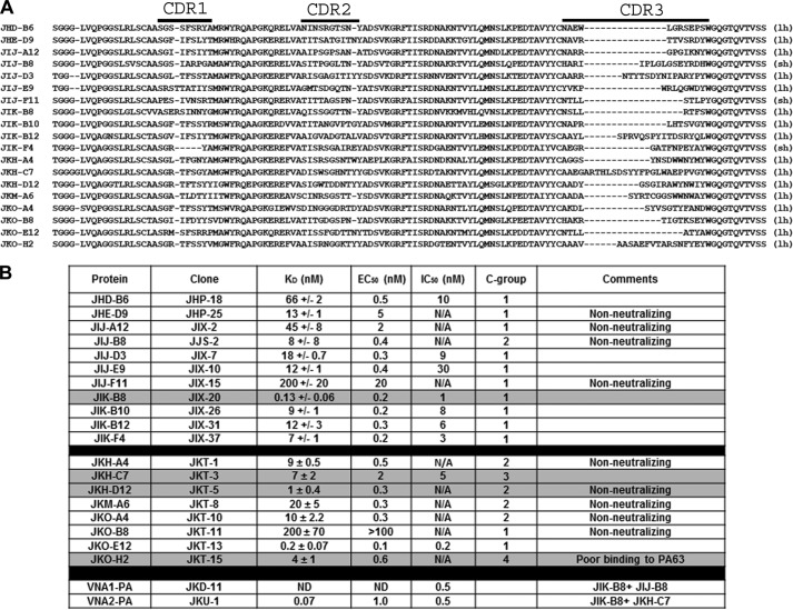
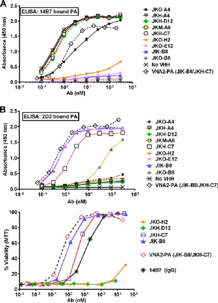
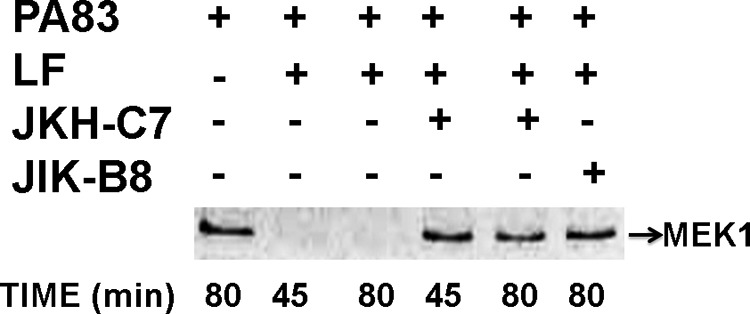
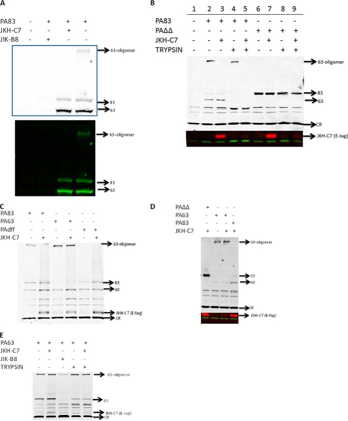
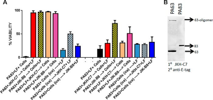
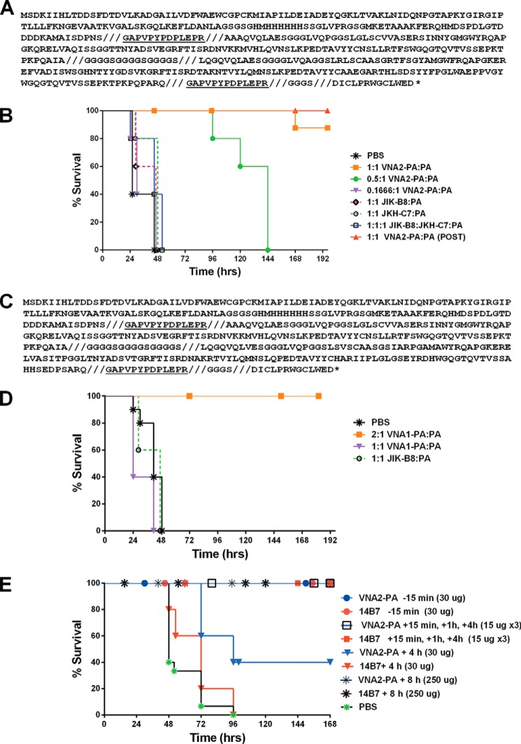
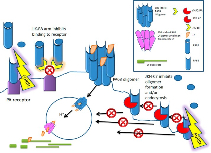
Anthrax disease is caused by a toxin consisting of protective antigen (PA), lethal factor, and edema factor. Antibodies against PA have been shown to be protective against the disease. Variable domains of camelid heavy chain-only antibodies (VHHs) with affinity for PA were obtained from immunized alpacas and screened for anthrax neutralizing activity in macrophage toxicity assays. Two classes of neutralizing VHHs were identified recognizing distinct, non-overlapping epitopes. One class recognizes domain 4 of PA at a well characterized neutralizing site through which PA binds to its cellular receptor. A second neutralizing VHH (JKH-C7) recognizes a novel epitope. This antibody inhibits conversion of the PA oligomer from "pre-pore" to its SDS and heat-resistant "pore" conformation while not preventing cleavage of full-length 83-kDa PA (PA83) by cell surface proteases to its oligomer-competent 63-kDa form (PA63). The antibody prevents endocytosis of the cell surface-generated PA63 subunit but not preformed PA63 oligomers formed in solution. JKH-C7 and the receptor-blocking VHH class (JIK-B8) were expressed as a heterodimeric VHH-based neutralizing agent (VNA2-PA). This VNA displayed improved neutralizing potency in cell assays and protected mice from anthrax toxin challenge with much better efficacy than the separate component VHHs. The VNA protected virtually all mice when separately administered at a 1:1 ratio to toxin and protected mice against Bacillus anthracis spore infection. Thus, our studies show the potential of VNAs as anthrax therapeutics. Due to their simple and stable nature, VNAs should be amenable to genetic delivery or administration via respiratory routes.

Keywords: Anthrax Toxin; Antibody; Antibody Engineering; Receptor; Toxin.


Figures

FIGURE 1.
FIGURE 1.
PA binding VHHs identified in this work. A, amino acid sequences of unique VHHs selected for binding to anthrax PA are shown. Sequences shown begin within framework 1 at the site of the primer binding employed in coding sequence DNA amplification from the immune alpaca cDNA (43) and continue through the end of framework 4. The parentheses at the end indicate whether the VHH contains a long hinge (lh) or a short hinge (sh). The three complementarity determining regions (CDR) are indicated at the top. B, table of the VHH names and binding properties. The first 11 VHHs were obtained by panning on PA83 bound to plastic, and the KD values for these VHHs were assessed by SPR-Proteon. The second group of nine VHHs were obtained by panning on 14B7-bound PA83 and the KD values assessed by SPR-Biacore. The KD for JIK-B8, obtained as an internal reference for SPR-Biacore group, was 1 ± 0.7. EC50 values were assessed by dilution ELISAs. IC50 values were assessed by toxin neutralization assays on macrophages and competition groups (C-groups) by competition ELISA. N/A refers to antibodies that did not neutralize toxin and thus have no measurable IC50.
FIGURE 2.
FIGURE 2.
Competition ELISAs and neutralization assays. A and B, 14B7- or 2D3-captured PA was bound to plates, and varying concentration of each VHH were then added and their binding assessed with an HRP-conjugated anti-His6 or E-tag antibody (Ab) using standard ELISA protocols. C, representative neutralization assays for the four VHHs representing the four competition groups, a heteromultimer of two neutralizing VHHs, and mAb 14B7 are shown. All assays are representative of at least two separate experiments.
FIGURE 3.
FIGURE 3.
JKH-C7 and JIK-B8 inhibit LF-mediated MEK-1 cleavage in macrophages. RAW264.7 cells were treated with PA83 + LF (200 ng/ml each) for the shown amounts of time or were first preincubated with 1 μg/ml VHHs for 30 min before the addition to cells for the indicated times. Cell lysates were subjected to Western blotting with an antibody that reacts to the MEK-1 N-terminal epitope that is lost after cleavage by LF. Western blots are representative of at least two separate experiments.
FIGURE 4.
FIGURE 4.
JKH-C7 does not prevent PA binding or cleavage and inhibits oligomer endocytosis. Various PA83, mutant PA proteins, or PA63 (1 μg/ml) were preincubated with antibodies (VHH:toxin, 4:1) in serum-free DMEM for 30 min to 1 h before the addition to CHO WTP4 cells for 60 min. Cells were either lysed directly in radioimmune precipitation assay buffer without trypsinization or trypsinized as described under “Experimental Procedures” to remove all cell surface proteins before lysis. Western blotting was performed with anti-PA polyclonal (1:5000), and in B–E, the blots were re-probed with goat anti-E-tag antibody (1:1000) to detect the JKH-C7 VHH in or on cells. IR-dye-conjugated secondary antibodies of different wavelengths were used to detect the PA and anti-E-tag antibodies in panels B and D where the re-probed section of the blot is shown as a separate panel. In panels C and E a single wavelength IR-dye secondary was used for detection of both primary antibodies, and the whole re-probed blot is shown. In panel A both a color and black and white image for the same blot probed with a single IR-dye secondary is shown for better identification of oligomer and lanes. CR stands for “cross-reactive” band, which serves as a nonspecific equal loading control. Western blots are representative of at least two separate experiments.
FIGURE 5.
FIGURE 5.
JKH-C7 does not inhibit preformed PA63 oligomer function. A, in columns 1–5 and 9–13, PA83 or PA63 (1 μg/ml) with or without LF (1 μg/ml) were incubated with VHH (4:1 VHH:toxin) or with medium (columns 1 and 9) for 1 h before the addition to cells. If LF was not present initially, it was present on cells (1 μg/ml) when the antibody-bound PA83 of PA63 was added. Cells were washed after 1 h of toxin binding and incubated overnight in complete medium before viability testing. In parallel plates, the PA83 or PA63 (1 μg/ml) was first bound to cells on ice for 1 h and washed with cold medium before the addition of the first VHH, transfer to 37 °C, and then LF in same amounts described above. Cells stayed at 37 °C for overnight incubation followed by viability assessment. The viability was calculated relative to untreated controls. Data presented in this graph are a representative experiment in which each column is the average of three replicate wells. B, purified PA63 or PA83 were boiled in SDS-loading buffer before SDS-PAGE and Western blotting in a triple sandwich using JKH-C7 as primary antibody, the goat anti-E-tag antibody as a secondary antibody and an IRDye-conjugated anti-goat antibody to detect the E-tag antibody. Western blots are representative of at least two separate experiments.
FIGURE 6.
FIGURE 6.
VNA amino acid sequence and antitoxin protection in animal models. A, VNA2-PA translation product sequence. Shown is the protein sequence of the entire VNA2-PA translation product expressed in E. coli. The VNA contains an N-terminal thioredoxin fusion partner and hexahistidine encoded by the pET32b expression vector. The VHH sequences are flanked by two E-tag peptides (underlined) and separated by an unstructured spacer ((GGGGS)3). A 14-amino acid albumin binding peptide (ABP), DICLPRWGCLEWED (36), is at the carboxyl end, separated from the second E-tag by a GGGGS spacer. For clarity, the eight defined protein segments are separated by slashed (///). B, Balb/cJ mice (n = 5/group, except PBS control group, n = 10) were injected intravenous with antibody at the indicated molar ratios (Ab:toxin) 10 min before injection with LT (45 μg for each toxin component, intravenous) except a single group marked (POST) that received antibody 2 h after toxin was administered. Control groups received PBS instead of antibody. Animals were monitored for 10 days post-intoxication for signs of malaise and survival. C, protein sequence of the entire VNA1-PA translation product with same specifications described in panel A. This VNA is a heterodimer of the neutralizing VHH JIK-B8 and the high affinity non-neutralizing antibody JIJ-B8. D, Balb/cJ mice were injected intravenously with antibody at the indicated molar ratios (Ab:toxin) 10 min before injection with LT (45 μg for each toxin component, intravenous). Control groups received PBS instead of antibody. Animals were monitored for 10 days post infection for signs of malaise and survival. E, C57BL/6J mice (n = 5/group, except PBS controls, n = 15) were treated with heterodimeric VNA2-PA (subcutaneously) at the indicated times and doses before or after spore infection (2 × 10⁷ spores, also subcutaneously at the distal site). Control mice were treated with PBS at 15 min, 1 h, and 4 h post infection (n = 5) or at 5 min (n = 5) and 8 h (n = 5) post infection. Neutralizing mAb 14B7 was used as a positive control in these studies. Mice were monitored for survival and signs of malaise for 10 days.
FIGURE 7.
FIGURE 7.
Model for mechanism of action of two VHHs that make up neutralizing VNA2-PA: PA83 normally binds to cellular receptors and is cleaved by cellular furin to form PA63. PA63 monomers immediately oligomerize to form the binding sites for LF. The oligomer complex is endocytosed (with or without LF cargo) and upon arrival in the acidic endosomal environment alters in conformation to form a pore through which LF can be translocated to the cytosol where it cleaves its substrates. VNA2-PA, which is a heterodimer of JIK-B8 and JKH-C7, inhibits this process through two mechanisms. The JIK-B8 arm binds to the receptor binding domain of PA83 and inhibits binding of the toxin to its receptor. The JKH-C7 arm binds to a conformational epitope on PA83 that does not prevent cleavage of the toxin to PA63 form but prevents oligomerization of PA63 at the cell surface or its endocytosis into the cell.

References

    1. Liu S., Moayeri M., Leppla S. H. (2014) Anthrax lethal and edema toxins in anthrax pathogenesis. Trends Microbiol. 22, 317–325 - PMC - PubMed
    1. Moayeri M., Leppla S. H. (2009) Cellular and systemic effects of anthrax lethal toxin and edema toxin. Mol. Aspects Med. 30, 439–455 - PMC - PubMed
    1. Chen Z., Moayeri M., Purcell R. (2011) Monoclonal antibody therapies against anthrax. Toxins 3, 1004–1019 - PMC - PubMed
    1. Kaur M., Singh S., Bhatnagar R. (2013) Anthrax vaccines: present status and future prospects. Expert Rev. Vaccines 12, 955–970 - PubMed
    1. Muyldermans S. (2013) Nanobodies: natural single-domain antibodies. Annu. Rev. Biochem. 82, 775–797 - PubMed

Publication types

MeSH terms