Review
. 2015 Jan 15;142(2):242-57.
doi: 10.1242/dev.105445.

Establishing neural crest identity: a gene regulatory recipe

Affiliations
Review

Establishing neural crest identity: a gene regulatory recipe

Marcos Simões-Costa et al. Development. .

Abstract

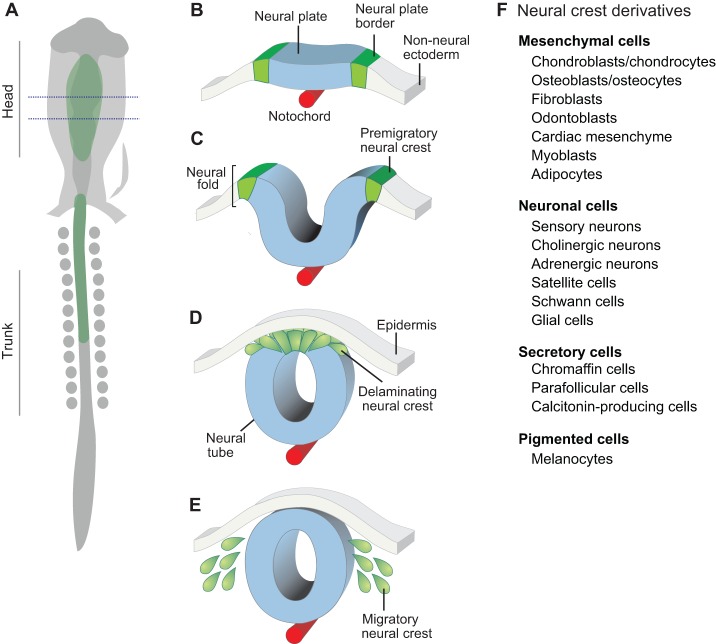
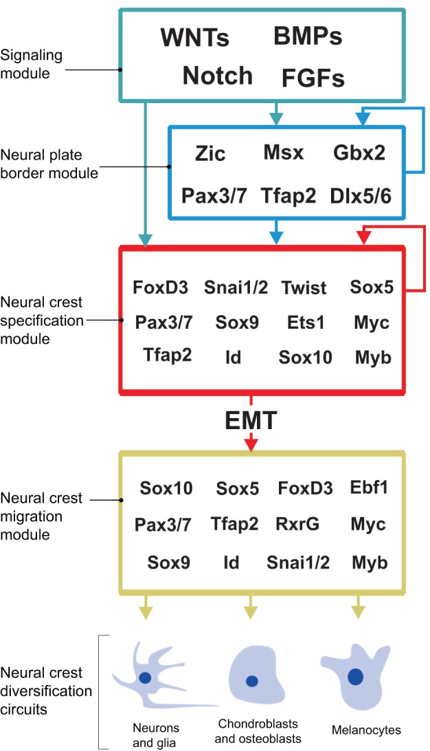
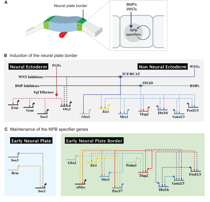
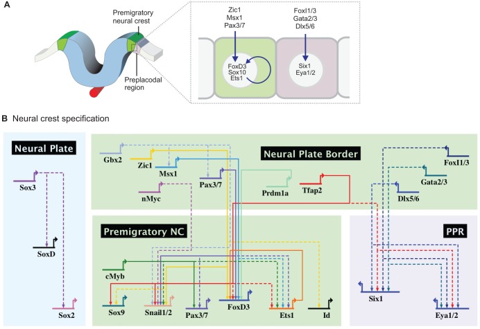
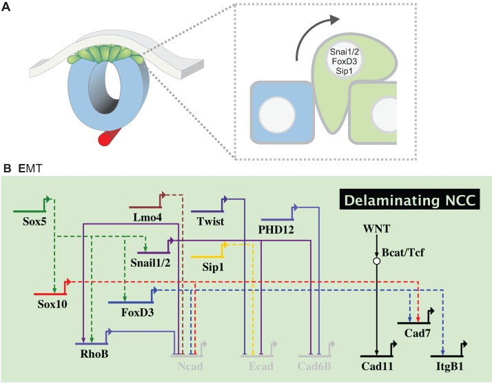
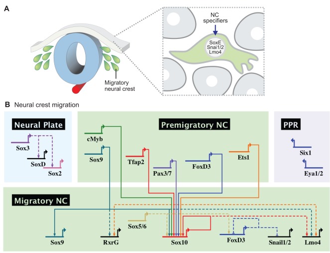
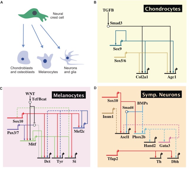
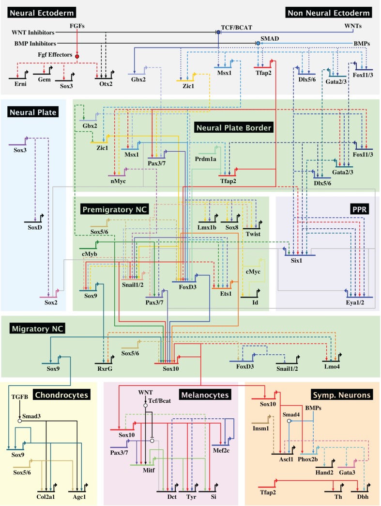
The neural crest is a stem/progenitor cell population that contributes to a wide variety of derivatives, including sensory and autonomic ganglia, cartilage and bone of the face and pigment cells of the skin. Unique to vertebrate embryos, it has served as an excellent model system for the study of cell behavior and identity owing to its multipotency, motility and ability to form a broad array of cell types. Neural crest development is thought to be controlled by a suite of transcriptional and epigenetic inputs arranged hierarchically in a gene regulatory network. Here, we examine neural crest development from a gene regulatory perspective and discuss how the underlying genetic circuitry results in the features that define this unique cell population.

Keywords: Gene regulation; Migration; Neural crest; Neural plate border; Signaling; Transcription factors.


Figures

Fig. 1.
Fig. 1.
The neural crest is a multipotent cell population. (A) Schematic dorsal view of a ten-somite stage chicken embryo, showing the neural crest (green) in the vicinity of the midline. The dotted lines delimit the embryonic region represented in cross-section (B-E). (B) Development of the neural crest begins at the gastrula stage, with the specification of the neural plate border at the edges of the neural plate. (C) As the neural plate closes to form the neural tube, the neural crest progenitors are specified in the dorsal part of the neural folds. (D) After specification, the neural crest cells undergo EMT and delaminate from the neural tube. (E) Migratory neural crest cells follow stereotypical pathways to diverse destinations, where they will give rise to distinct derivatives. (F) The neural crest is multipotent and has the capacity to give rise to diverse cell types, including cells of mesenchymal, neuronal, secretory and pigmented identity.
Fig. 2.
Fig. 2.
A GRN controls neural crest formation. Outline of the GRN controlling neural crest development. Different inductive signals pattern the embryonic ectoderm and induce the expression of neural plate border specifier genes, which define the neural plate border territory. These genes engage in mutual positive regulation and also drive neural crest specification by activating the neural crest specifier genes. The neural crest specification program results in the activation of the EMT machinery that allows the neural crest cells to become migratory. The migratory neural crest cells express a set of regulators that endow them with motility and the ability to initiate different differentiation programs.
Fig. 3.
Fig. 3.
GRN module controlling the formation of the neural plate border. (A) The action of signaling systems such the BMP and WNT signaling pathways results in activation of the neural plate border (NPB) genes in the margins of the neural plate. (B) In the neural ectoderm, FGF signaling drives neural induction by activating proneural genes. In an intermediary territory located between the neural and non-neural ectoderm, WNTs and BMPs activate a number of transcription factors dubbed neural plate border specifiers. Activity of WNTs and BMPs is hampered in the neural ectoderm by a number of inhibitory molecules produced by these cells. (C) While SoxB1 genes (Sox2 and Sox3) are expressed in the early neural plate, the neural plate specifier transcription factors engage in mutual positive regulation, which stabilizes the neural plate border regulatory state. Direct interactions are indicated with solid lines, whereas dashed lines show possible direct interactions inferred from gain- and loss-of-function studies.
Fig. 4.
Fig. 4.
Gene regulatory interactions controlling specification of the neural crest. (A) As the neural plate folds, the neural plate border specifier genes Zic1, Msx1 and Pax3/7 activate the expression of neural crest specifier genes FoxD3, Sox10 and Ets1. Lateral to the neural crest cells, preplacodal cells express a distinct set of transcription factors. (B) Expression of neural crest specifiers in the premigratory neural crest (NC) territory is mediated by regulators in the medial regions of the neural plate border. The neighboring preplacodal region (PPR) is defined by expression of Six1 and Eye1/2, which identify the progenitor field that will give rise to cranial placodes. SoxD and SoxB1 genes are expressed in the neural plate.
Fig. 5.
Fig. 5.
Regulatory module responsible for neural crest EMT. Activity of Snai1/2, FoxD3, Twist and Zeb2 (Sip1) mediate changes in cell-cell interaction that allow for delamination and dispersion of neural crest cells (NCC). Repression of the epithelial cadherins Ncad, Ecad and Cad6b and activation of the type 2 cadherins Cad11 and Cad7 allow for changes in cell adhesion and EMT.
Fig. 6.
Fig. 6.
Neural crest specifiers drive the transition to the migratory neural crest regulatory state. (A) Action of neural crest specifiers transforms the identity of the neural crest as it becomes a migratory cell population. (B) Neural crest specifiers (including Sox9, cMyb and Ets1) cooperate to drive the expression of genes that are strongly upregulated in the migratory neural crest, such as Sox10. This transcription factor positively regulates itself, which results in maintenance of Sox10 expression as the neural crest cells migrate throughout the embryo to give rise to different derivatives.
Fig. 7.
Fig. 7.
Subcircuits controlling neural crest diversification. (A) Neural crest cells give rise to distinct derivatives depending on their migratory pathways and external cues. (B) Differentiation of neural crest to chondrocytes depends on the activity of Sox9 and Sox5/6, which cooperate with TGFβ signaling to directly activate the cartilage differentiation genes Col2a1 and Agc1. (C) Differentiation to melanocytes is centered on a Sox10-Mitf feed-forward loop. These transcription factors cooperate to directly activate Dct, Tyr and Si, which encode melanogenic enzymes. (D) BMP signaling drives the differentiation of neural crest cells to sympathetic neurons through activation of Ascl1 and Phox2b, which in turn activate a circuit controlling differentiation genes such as tyrosine hydroxylase (Th) and dopamine β-hydroxylase (Dbh).
Fig. 8.
Fig. 8.
The GRN controlling neural crest development. Regulatory information obtained from different vertebrate model organisms has been assembled to produce a GRN that controls the formation and diversification of the neural crest. Direct interactions, supported by cis-regulatory analysis, are represented by solid lines, whereas dashed lines indicate interactions inferred from gene perturbation analysis.

References

    1. Agarwal P., Verzi M. P., Nguyen T., Hu J., Ehlers M. L., McCulley D. J., Xu S.-M., Dodou E., Anderson J. P., Wei M. L. et al. (2011). The MADS box transcription factor MEF2C regulates melanocyte development and is a direct transcriptional target and partner of SOX10. Development 138, 2555-2565 10.1242/dev.056804 - DOI - PMC - PubMed
    1. Aruga J. (2004). The role of Zic genes in neural development. Mol. Cell. Neurosci. 26, 205-221 10.1016/j.mcn.2004.01.004 - DOI - PubMed
    1. Attanasio C., Nord A. S., Zhu Y., Blow M. J., Li Z., Liberton D. K., Morrison H., Plajzer-Frick I., Holt A., Hosseini R. et al. (2013). Fine tuning of craniofacial morphology by distant-acting enhancers. Science 342, 1241006 10.1126/science.1241006 - DOI - PMC - PubMed
    1. Aybar M. J., Nieto M. A. and Mayor R. (2003). Snail precedes slug in the genetic cascade required for the specification and migration of the Xenopus neural crest. Development 130, 483-494 10.1242/dev.00238 - DOI - PubMed
    1. Bae C.-J., Park B.-Y., Lee Y.-H., Tobias J. W., Hong C.-S. and Saint-Jeannet J.-P. (2014). Identification of Pax3 and Zic1 targets in the developing neural crest. Dev. Biol. 386, 473-483 10.1016/j.ydbio.2013.12.011 - DOI - PMC - PubMed

Publication types

MeSH terms