. 2015 Feb 1;142(3):438-43.
doi: 10.1242/dev.113571. Epub 2015 Jan 6.

Tetraspanin CD9 and ectonucleotidase CD73 identify an osteochondroprogenitor population with elevated osteogenic properties

Affiliations

Tetraspanin CD9 and ectonucleotidase CD73 identify an osteochondroprogenitor population with elevated osteogenic properties

Anju Singh et al. Development. .

Abstract

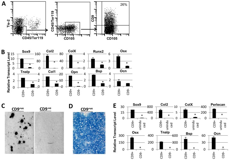
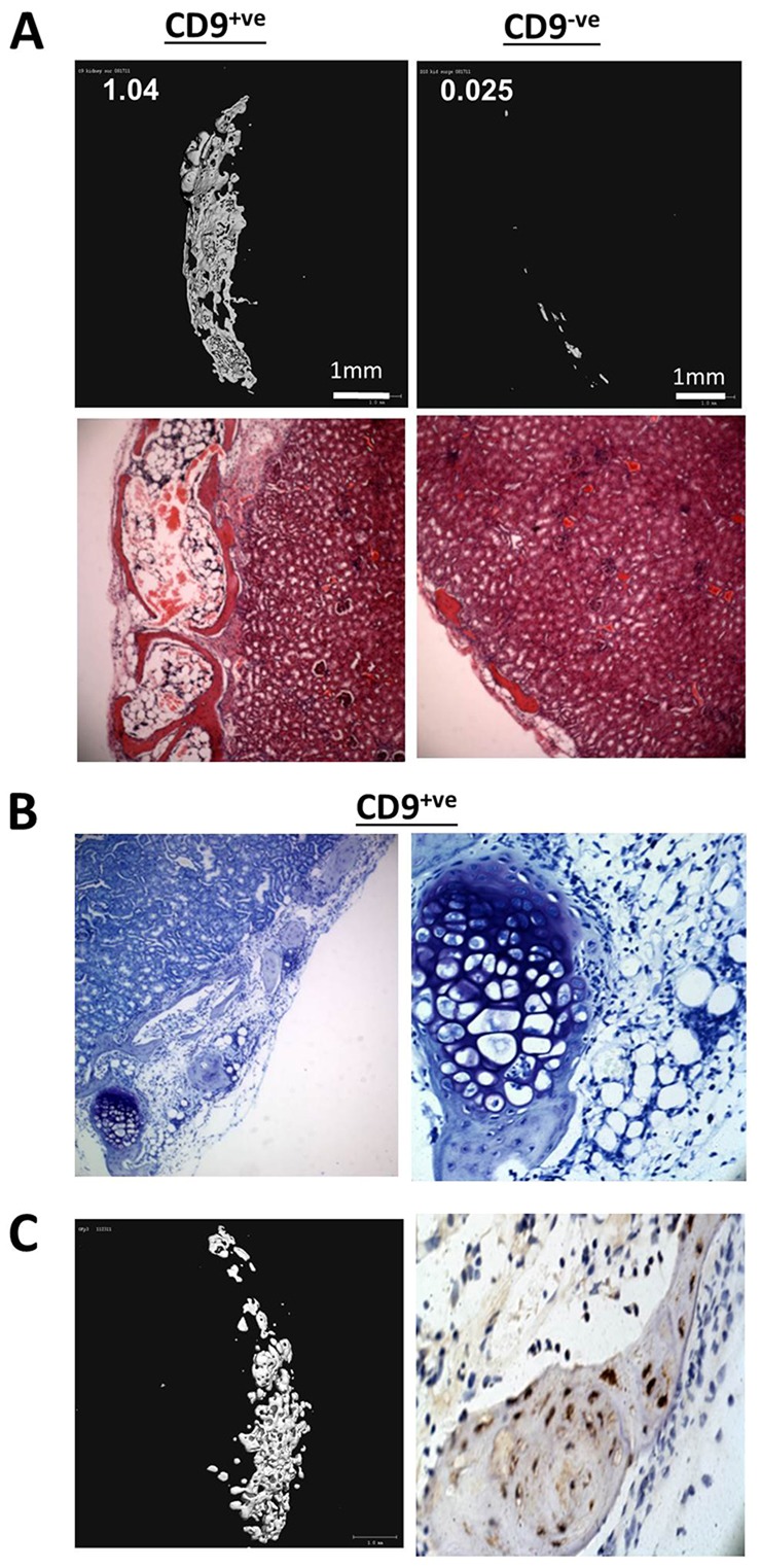
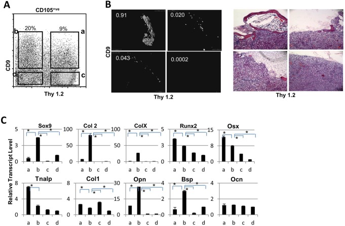
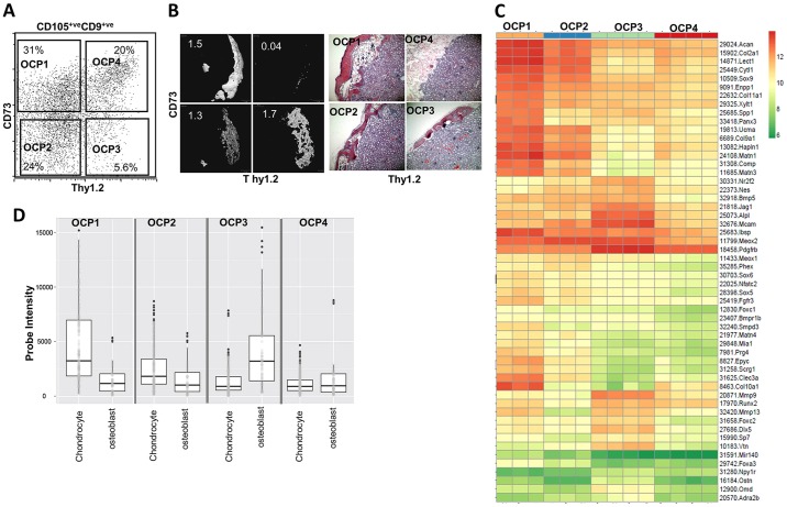
Cell-based bone regeneration strategies offer promise for traumatic bone injuries, congenital defects, non-union fractures and other skeletal pathologies. Postnatal bone remodeling and fracture healing provide evidence that an osteochondroprogenitor cell is present in adult life that can differentiate to remodel or repair the fractured bone. However, cell-based skeletal repair in the clinic is still in its infancy, mostly due to poor characterization of progenitor cells and lack of knowledge about their in vivo behavior. Here, we took a combined approach of high-throughput screening, flow-based cell sorting and in vivo transplantation to isolate markers that identify osteochondroprogenitor cells. We show that the presence of tetraspanin CD9 enriches for osteochondroprogenitors within CD105(+) mesenchymal cells and that these cells readily form bone upon transplantation. In addition, we have used Thy1.2 and the ectonucleotidase CD73 to identify subsets within the CD9(+) population that lead to endochondral or intramembranous-like bone formation. Utilization of this unique cell surface phenotype to enrich for osteochondroprogenitor cells will allow for further characterization of the molecular mechanisms that regulate their osteogenic properties.

Keywords: Osteoblast; Osteochondroprogenitor; Skeletal stem cell.


Figures

Fig. 1.
Fig. 1.
Identification of CD9 as a marker for osteochondroprogenitors. (A) Representative flow plots of mice embryonic limb suspension at E16.5 stained with anti-CD45, anti-Ter119, anti-Tie2, anti-CD105 and anti-CD9. The dot plot on the right is pre-gated on CD45Tie2Ter119CD105+ cells. (B) qPCR expression of the indicated transcripts in sorted CD9+ or CD9 from E16.5 fetal limbs. (C) Von Kossa staining of sorted CD9+ or CD9 cells from E16.5 fetal bones after 15 days in osteoblast differentiation media. (D) Toluidine Blue staining of section of chondrocyte pellet from sorted CD9+ cells after 2 weeks in chondrocyte differentiation media. (E) qPCR expression of the indicated transcripts in chondrocyte pellets from sorted E16.5 CD9+ cells after 2 weeks in chondrocyte-inducing conditions (top panel) or sorted E16.5 CD9+ or CD9 after 2 weeks in osteoblast-differentiating conditions (bottom panel). *P<0.05; Student's t-test.
Fig. 2.
Fig. 2.
CD9+ osteochondroprogenitors give rise to bone and cartilage in vivo. (A) Representative 3D micro-CT images (upper panel) and H&E staining (lower panel) of kidneys transplanted with 20,000 sorted CD9+ or CD9 cells. Kidneys were harvested 6 weeks post cell transfer and scanned using a micro-CT machine. The region of interest (ROI) was defined to include the entire kidney, and 2D image stacks were visually inspected to ensure that all heterotopic bone was included within the ROI. The numbers represent total bone volume in the ROI, calculated using built-in Scanco software. (B) Toluidine Blue staining of kidney sections 4 weeks after transplantation with 20,000 sorted CD9+ or CD9 cells. (C) 20,000 sorted CD9+ cells were transferred from GFP transgenic mice to non-transgenic mice. Kidneys were harvested 4 weeks post transplantation and were analyzed by micro-CT (left), or sections were immunostained against GFP (right).
Fig. 3.
Fig. 3.
The cell surface marker Thy1.2 further sub-characterizes CD9+ osteochondroprogenitors. (A) Representative flow plot of mice embryonic limb suspension at E16.5 stained with anti-CD45, anti-Ter119, anti-Tie2, anti-CD105, anti-Thy1.2 and anti-CD9. The plot is pre-gated on CD105+ cells. (B) Representative 3D micro-CT images (left) and H&E staining (right) of kidneys 6 weeks post transplantation with 20,000 sorted CD9+Thy1.2+, CD9+Thy1.2, CD9Thy1.2+ or CD9Thy1.2 E16.5 fetal bone cells. Numbers represent total bone volume calculated using micro-CT. (C) qPCR expression of indicated transcripts from sorted CD9+Thy1.2+ (a), CD9+Thy1.2 (b), CD9Thy1.2+ (c) or CD9Thy1.2 (d) E16.5 fetal bone suspension. *P<0.05; Student's t-test.
Fig. 4.
Fig. 4.
The ecto-5′-nucleotidase CD73 further sub-characterizes the CD9+ osteochondroprogenitors. (A) Dot plot showing staining with anti-CD73 and anti-Thy1.2 at E16.5. The plot is pre-gated on CD105+CD9+ cells. (B) Representative 3D micro-CT images and H&E staining of kidneys transplanted with 20,000 sorted OCP1 (CD73+Thy1.2), OCP2 (CD73Thy1.2), OCP3 (CD73Thy1.2+) and OCP4 (CD73+Thy1.2+) fetal bone suspension at E16.5. Kidneys were harvested 6 weeks post cell transfer. (C) Heat map showing genes differentially expressed between OCP1, OCP2, OCP3 and OCP4 fetal bone cells at E16.5. (D) Box plots demonstrating gene intensity expression between different populations. Differentially expressed genes were identified using limma and manually curated for any association with chondrocytes or osteoblasts.

Similar articles

Cited by

References

    1. Bakker A. D. and Klein-Nulend J. (2012). Osteoblast isolation from murine calvaria and long bones. Methods Mol. Biol. 816, 19-29 10.1007/978-1-61779-415-5_2 - DOI - PubMed
    1. Bianco P., Robey P. G. and Simmons P. J. (2008). Mesenchymal stem cells: revisiting history, concepts, and assays. Cell Stem Cell 2, 313-319 10.1016/j.stem.2008.03.002 - DOI - PMC - PubMed
    1. Bianco P., Robey P. G., Saggio I. and Riminucci M. (2010). “Mesenchymal” stem cells in human bone marrow (skeletal stem cells): a critical discussion of their nature, identity, and significance in incurable skeletal disease. Hum. Gene Ther. 21, 1057-1066 10.1089/hum.2010.136 - DOI - PMC - PubMed
    1. Boucheix C. and Rubinstein E. (2001). Tetraspanins. Cell. Mol. Life Sci. 58, 1189-1205 10.1007/PL00000933 - DOI - PMC - PubMed
    1. Chan C. K. F., Chen C.-C., Luppen C. A., Kim J.-B., DeBoer A. T., Wei K., Helms J. A., Kuo C. J., Kraft D. L. and Weissman I. L. (2009). Endochondral ossification is required for haematopoietic stem-cell niche formation. Nature 457, 490-494 10.1038/nature07547 - DOI - PMC - PubMed

Publication types

MeSH terms

Associated data