. 2015 Jan 7:6:5935.
doi: 10.1038/ncomms6935.

Crystallographic and spectroscopic snapshots reveal a dehydrogenase in action

Affiliations

Crystallographic and spectroscopic snapshots reveal a dehydrogenase in action

Lu Huo et al. Nat Commun. .

Abstract

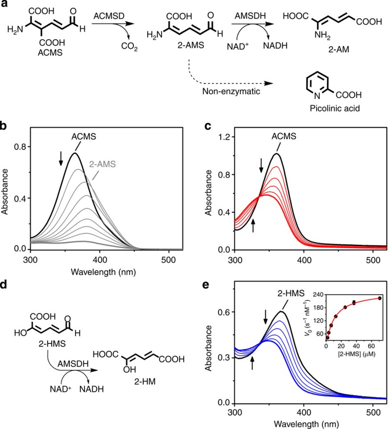
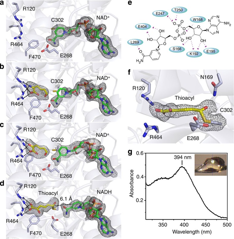
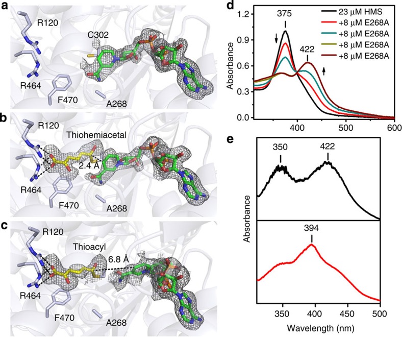
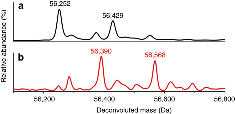
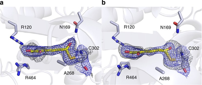
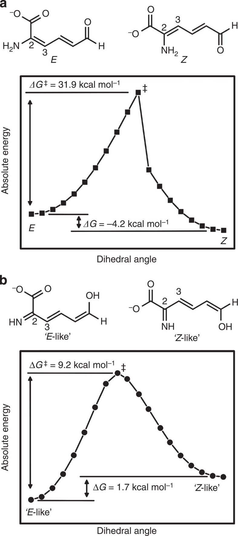
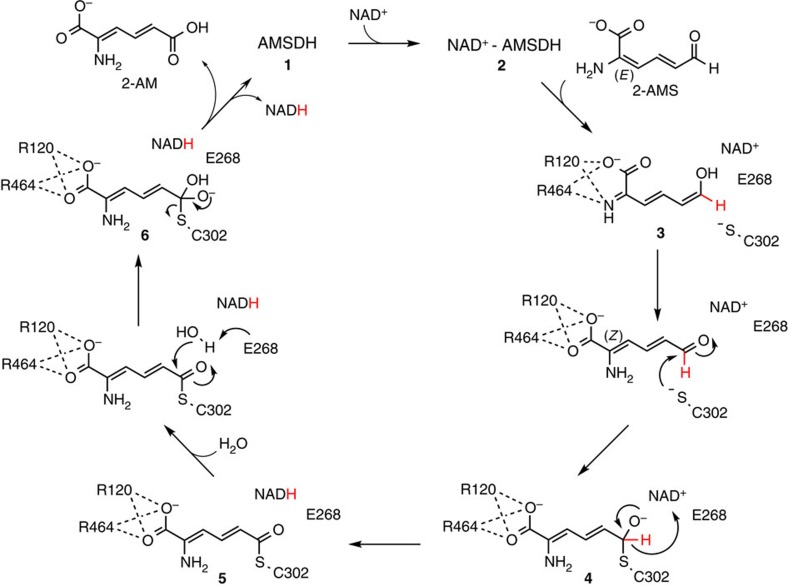
Aldehydes are ubiquitous intermediates in metabolic pathways and their innate reactivity can often make them quite unstable. There are several aldehydic intermediates in the metabolic pathway for tryptophan degradation that can decay into neuroactive compounds that have been associated with numerous neurological diseases. An enzyme of this pathway, 2-aminomuconate-6-semialdehyde dehydrogenase, is responsible for 'disarming' the final aldehydic intermediate. Here we show the crystal structures of a bacterial analogue enzyme in five catalytically relevant forms: resting state, one binary and two ternary complexes, and a covalent, thioacyl intermediate. We also report the crystal structures of a tetrahedral, thiohemiacetal intermediate, a thioacyl intermediate and an NAD(+)-bound complex from an active site mutant. These covalent intermediates are characterized by single-crystal and solution-state electronic absorption spectroscopy. The crystal structures reveal that the substrate undergoes an E/Z isomerization at the enzyme active site before an sp(3)-to-sp(2) transition during enzyme-mediated oxidation.


Figures

Figure 1
Figure 1. Activity of AMSDH.
(a) Reaction scheme showing the enzymatic generation of 2-AMS, the reaction catalysed by AMSDH, and the competing non-enzymatic decay of 2-AMS to picolinic acid. (b) Representative assay showing the ACMSD (1 μM)-catalysed conversion of ACMS (λmax 360 nm) to 2-AMS (λmax 380 nm), which decays to picolinic acid (transparent). (c) Coupled-enzyme assay in which AMSDH (200 nM) oxidizes 2-AMS, produced in situ as shown in b in 50 s, to 2-AM (λmax 325 nm). (d) Reaction scheme showing 2-HMS oxidation by AMSDH. (e) Representative assay showing the activity of AMSDH (200 nM) on 2-HMS (λmax 375 nm) in 50 s. The inset is a Michaelis–Menten plot.
Figure 2
Figure 2. Crystal structures of wild-type AMSDH and single-crystal electronic absorption spectrum of a catalytic intermediate.
AMSDH was co-crystallized with NAD+ to give AMSDH-NAD+ binary complex crystals that were used for soaking experiments. (a) Active site structure of the binary AMSDH-NAD+ complex, (b) the ternary complex of AMSDH-NAD+ crystals soaked with 2-AMS for 5 min before flash cooling, (c) the ternary complex of AMSDH-NAD+ soaked with 2-HMS for 10 min before flash cooling, (d) the trapped thioacyl, NADH-bound intermediate obtained by soaking AMSDH-NAD+ crystals with 2-HMS for 40 min before flash cooling. (e) Two-dimensional interaction diagram for NAD+ binding. (f) Close-up of the thioacyl intermediate in d. (g) Single-crystal electronic absorption spectrum of d. Protein backbone and residues are shown as light blue cartoons and sticks, respectively. The substrates and intermediate are shown as yellow sticks, and NAD+ and NADH are shown as green sticks. The omit map for ligands is contoured to 2.0 σ and shown as a grey mesh.
Figure 3
Figure 3. Crystal structures of the E268A mutant and its solution and single-crystal electronic absorption spectra.
(a) Structure of the active site of the co-crystallized E268A-NAD+ binary complex, (b) a thiohemiacetal intermediate obtained by soaking the E268A-NAD+ crystals with 2-HMS for 30 min before flash cooling and (c) a thioacyl intermediate obtained by soaking the E268A-NAD+ crystals with 2-HMS for 180 min before flash cooling. (d) Solution electronic absorption spectra of a titration of 2-HMS with E268A. (e) Single-crystal electronic absorption spectrum of the intermediate in b (top panel) and single-crystal electronic absorption spectrum of the intermediate in c (bottom panel). Protein backbone and residues are shown as light blue cartoons and sticks, respectively. The substrate and intermediate are shown as yellow sticks, and NAD+ and NADH are shown as green sticks. The omit map for ligands is contoured to 2.0 σ and shown as a grey mesh.
Figure 4
Figure 4. Deconvoluted positive-mode electrospray ionization mass spectra of as-isolated E268A (a) and 2-HMS treated-E268A (b).
The two major components are labelled with their respective molecular weights.
Figure 5
Figure 5. Crystal structures of two distinct catalytic intermediates.
(a) Electron density map of the thiohemiacetal intermediate obtained from E268A-NAD+ crystal soaked with 2-HMS for 30 min. (b) Electron density map of the thioacyl intermediate obtained from E268A-NAD+ crystal soaked with 2-HMS for 180 min. The 2FoFc electron density map for ligands and Cys302 is contoured to 1.0 σ and shown as a blue mesh. The omit map for ligands and Cys302 is contoured to 2.0 σ and shown as a gray mesh.
Figure 6
Figure 6. Free energy profiles for the rotation about the 2–3 bond of 2-AMS in its (a) enamine and (b) imine form, respectively.
DFT calculations were performed at the B3LYP/6-31G*+ level of theory. The dihedral angle about the 2–3 bond was restrained in 10° increments and the structures were optimized at each point.
Figure 7
Figure 7. Proposed catalytic mechanism for the oxidation of 2-AMS by AMSDH.
The primary substrate (2E, 4E)-2-aminomuconate-semialdehyde binds to the enzyme in its imine tautomer to form the ternary complex (3). An isomerization and attack by cysteine on the aldehydic carbon form the (2Z, 4E)-2-aminomuconate-thiohemiacetal adduct (4). AMSDH-mediated oxidation of 4 concomitant with reduction of NAD+ to NADH follows, generating a thioacyl-enzyme intermediate (5). Both 4 and 5 are the catalytic intermediates covalently attached to the enzyme. Hydrolysis of 5 then allows the release of the products 2-AM and NADH, restoring the ligand-free enzyme for the next catalytic cycle.

References

    1. Keszthelyi D., Troost F. J. & Masclee A. A. Understanding the role of tryptophan and serotonin metabolism in gastrointestinal function. Neurogastroenterol. Motil. 21, 1239–1249 (2009). - PubMed
    1. Myint A. M. et al.. Kynurenine pathway in major depression: evidence of impaired neuroprotection. J. Affect. Disord. 98, 143–151 (2007). - PubMed
    1. Ogawa T. et al.. Kynurenine pathway abnormalities in Parkinson’s disease. Neurology 42, 1702–1706 (1992). - PubMed
    1. Guillemin G. J. et al.. Quinolinic acid in the pathogenesis of Alzheimer’s disease. Adv. Exp. Med. Biol. 527, 167–176 (2003). - PubMed
    1. Guidetti P. & Schwarcz R. 3-Hydroxykynurenine and quinolinate: pathogenic synergism in early grade Huntington’s disease? Adv. Exp. Med. Biol. 527, 137–145 (2003). - PubMed

Publication types

MeSH terms

Substances

LinkOut - more resources