. 2015 Jan 7;10(1):e0117154.
doi: 10.1371/journal.pone.0117154. eCollection 2015.

A bovine cell line that can be infected by natural sheep scrapie prions

Affiliations

A bovine cell line that can be infected by natural sheep scrapie prions

Anja M Oelschlegel et al. PLoS One. .

Erratum in

Abstract

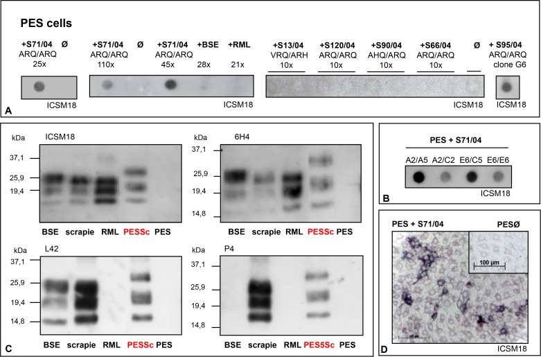
Cell culture systems represent a crucial part in basic prion research; yet, cell lines that are susceptible to prions, especially to field isolated prions that were not adapted to rodents, are very rare. The purpose of this study was to identify and characterize a cell line that was susceptible to ruminant-derived prions and to establish a stable prion infection within it. Based on species and tissue of origin as well as PrP expression rate, we pre-selected a total of 33 cell lines that were then challenged with natural and with mouse propagated BSE or scrapie inocula. Here, we report the successful infection of a non-transgenic bovine cell line, a sub-line of the bovine kidney cell line MDBK, with natural sheep scrapie prions. This cell line retained the scrapie infection for more than 200 passages. Selective cloning resulted in cell populations with increased accumulation of PrPres, although this treatment was not mandatory for retaining the infection. The infection remained stable, even under suboptimal culture conditions. The resulting infectivity of the cells was confirmed by mouse bioassay (Tgbov mice, Tgshp mice). We believe that PES cells used together with other prion permissive cell lines will prove a valuable tool for ongoing efforts to understand and defeat prions and prion diseases.


Conflict of interest statement

Competing Interests: The authors have declared that no competing interests exist.

Figures

Figure 1
Figure 1. Persistent infection of a bovine cell line (PES) with sheep scrapie prions.
A) PES cells were infected with the natural sheep scrapie isolates S71/04 and S95/04. Both isolates were derived from sheep carrying the ARQ/ARQ prion genotype. PrPres was detected by dot-blot analysis. Shown here are samples from three independent infections with S71/04, assayed 25, 110 or 45 cell passages after inoculation. S95/04-infected PESSc cells were used for cell cloning and shown here is PESSc clone G6. Uninfected control cells (ᴓ) or PES cells challenged with BSE or RML prions that were passaged 28 and 21 times, respectively, show no detectable PrPres signal. In addition the challenge with four other scrapie field isolates (S13/04, S120/04, S90/04 and S66/04) did not lead to PrPres propagation; shown here are samples after 10 cell passages post inocula application. B) Serial cloning of PESSc cells that had been infected with scrapie isolate S71/04 resulted in cell populations with different PrPres loads. Cell clones with stronger PrPres signals were selected for further cultivation. C) Detecting PrPres by western-blot PESSc prions were recognized by a panel of four different antibodies (ICSM18, 6H4, L42, P4) and show a higher molecular weight compared with BSE, scrapie and RML brain homogenate. D) The Cell-ELISA confirmed the infection of PES cells, showing PrPres positive single cells and cell accumulations. Uninfected PES cells are shown in the inlet.
Figure 2
Figure 2. Relative resistance of PESSc prions to ProteinaseK.
A) Cell lysates from PESSc cells, ScN2a cells and SMBRC040 cells were each divided into 13 aliquots. All aliquots, but one of each cell line, were subjected to ProteinaseK digestion (25 µg/ml; at 37°C). Every two hours the digestion of one aliquot per cell line was stopped with PMSF. PrPres was detected by dot-blot analysis (mAB ICSM18). In both murine cell lysates PrPres was detectable for up to 22 hours; in PESSc cell lysates PrPres was still detectable after 24 hours. B) Cell lysates from uninfected PES cells, PESSc cells and ScN2a cells were divided into six aliquots each, ProteinaseK was added at increasing concentrations (0 µg/ml, 25 µg/ml, 100 µg/ml, 250 µg/ml, 500 µg/ml and 1000 µg/ml), and the samples were incubated for one hour at 37°C. The digestion was terminated by adding PMSF. PrPres was detected by dot-blot analysis (mAB ICSM18). While 25 µg ProteinaseK/ml were sufficient to clear the control lysate from any detectable PrP, strong PrPres signals were detected in both prion infected samples up to 500 & 1000 µg ProteinaseK/ml.
Figure 3
Figure 3. Re-infection of „cured“ PESSc cells with different sheep scrapie field isolate.
PESSc cell clones A2/A5 and E6/C5, propagating ovine scrapie prions from the sheep scrapie field isolate S71/04, had been cured with the prion inhibitors Imatinib, Suramin and Pentosanpolysulfat until any detectable PrPres was cleared from the culture. Following that, the cells were cultured for 18 to 22 splits in the absence of the inhibitors and remained uninfected as monitored by dot-blot analysis. The cells were then subjected to infection with a second ovine scrapie field isolate, S95/04, and split 20 times at a ratio of 1:2. PrPres was detected by dot-blot analysis. mAB: ICSM18.
Figure 4
Figure 4. Verification of infectivity in PES cells by mouse bioassay.
A) Cell lysates of PESSc cells were inoculated into three groups of mice (six animals each group): Tgbov XV, Tgshp IX and C57/BL6. Tgbov XV mice succumbed to disease 245 ± 29 dpi, Tgshp IX mice died 444 ± 23 dpi and the first non-transgenic mouse was culled due to clinical signs 472 days post inoculation. The brain homogenates were tested by western-blot analysis for PrPres signals. B) Side by side western-blot analysis of PK digested PrPres derived from PESSc cells shows a higher molecular weight than PK digested PrPres from PESSc infected Tgbov XV or Tgshp IX mice. C) PES cells were inoculated with brain homogenate from Tgbov XV mouse 1. The cells were split 15 times at a ratio of 1:2 and were then assayed for PrPres by dot-blot analysis. Uninfected PES cells were assayed as control Ø. mAB: ICSM18.

References

    1. Prusiner SB (1982) Novel proteinaceous infectious particles cause scrapie. Science 216: 136–144. 10.1126/science.6801762 - DOI - PubMed
    1. Wang F, Wang X, Yuan CG, Ma J (2010) Generating a prion with bacterially expressed recombinant prion protein. Science 327: 1132–1135. 10.1126/science.1183748 - DOI - PMC - PubMed
    1. Castilla J, Saa P, Hetz C, Soto C (2005) In vitro generation of infectious scrapie prions. Cell 121: 195–206. 10.1016/j.cell.2005.02.011 - DOI - PubMed
    1. Saa P, Castilla J, Soto C (2006) Ultra-efficient replication of infectious prions by automated protein misfolding cyclic amplification. J Biol Chem 281: 35245–35252. 10.1074/jbc.M603964200 - DOI - PubMed
    1. Baron T (2002) Mouse models of prion disease transmission. Trends Mol Med 8: 495–500. 10.1016/S1471-4914(02)02416-4 - DOI - PubMed

MeSH terms