

Endodontic management of maxillary first molar with seven root canals diagnosed using Cone Beam Computed Tomography scanning
- PMID: 25565745
- PMCID: PMC4213875
- DOI: 10.4103/0975-962X.140837
Endodontic management of maxillary first molar with seven root canals diagnosed using Cone Beam Computed Tomography scanning
Abstract
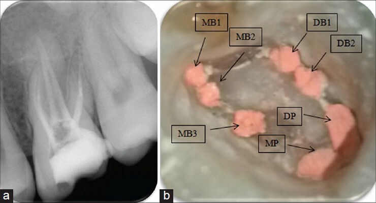
The main objective of root canal treatment is thorough cleaning and shaping of the entire pulp space and its complete filling with an inert filling material. A major cause of post-treatment disease is the inability to locate, debride or adequately fill all canals of the root canal system. The form, configuration, and number of root canals in the maxillary first molars have been discussed for more than half a century. Maxillary first molars commonly present with three roots and three canals, with a second mesiobuccal canal (MB2) also present. With the advent of improved magnification there are reports of multiple root canals in the maxillary first molars. Nonsurgical endodontic therapy of a left maxillary first molar with three roots and seven root canals was successfully performed under a dental operating microscope. The diagnosis of multiple root canals was confirmed with the help of Cone Beam Computed Tomography (CBCT) images.
Keywords: Aberrant canal anatomy; cone beam computed tomography; dental operating microscope; maxillary first molar.
Conflict of interest statement
Figures


Similar articles
-
The morphology of maxillary first and second molars analyzed by cone-beam computed tomography in a polish population.BMC Med Imaging. 2017 Dec 29;17(1):68. doi: 10.1186/s12880-017-0243-3. BMC Med Imaging. 2017. PMID: 29284426 Free PMC article.
-
[Root canal therapy of maxillary molars with atypical canals: A report of three cases].Beijing Da Xue Xue Bao Yi Xue Ban. 2024 Feb 18;56(1):190-195. doi: 10.19723/j.issn.1671-167X.2024.01.030. Beijing Da Xue Xue Bao Yi Xue Ban. 2024. PMID: 38318917 Free PMC article. Chinese.
-
A Cone-beam Computed Tomographic Study of Root and Canal Morphology of Maxillary First and Second Permanent Molars in a Thai Population.J Endod. 2018 Jan;44(1):56-61. doi: 10.1016/j.joen.2017.08.020. Epub 2017 Oct 20. J Endod. 2018. PMID: 29061352
-
Three root canals in the mesiobuccal root of maxillary molars: case reports and literature review.J Endod. 2014 Dec;40(12):2087-94. doi: 10.1016/j.joen.2014.07.034. Epub 2014 Oct 16. J Endod. 2014. PMID: 25443283 Review.
-
Evaluation of root canal morphology of human primary molars by using CBCT and comprehensive review of the literature.Acta Odontol Scand. 2016;74(4):250-8. doi: 10.3109/00016357.2015.1104721. Epub 2015 Nov 2. Acta Odontol Scand. 2016. PMID: 26523502 Review.
Cited by
-
Maxillary first molar with 7 root canals diagnosed using cone-beam computed tomography.Restor Dent Endod. 2017 Feb;42(1):60-64. doi: 10.5395/rde.2017.42.1.60. Epub 2016 Aug 29. Restor Dent Endod. 2017. PMID: 28194366 Free PMC article.
-
Detection and management of a complex canal configuration in mesiobuccal root of maxillary first molar using three dimensional imaging.J Int Soc Prev Community Dent. 2016 Apr;6(Suppl 1):S75-8. doi: 10.4103/2231-0762.181190. J Int Soc Prev Community Dent. 2016. PMID: 27195233 Free PMC article.
-
Comparison of apical debris extrusion of two rotary systems and one reciprocating system.J Conserv Dent. 2016 May-Jun;19(3):245-9. doi: 10.4103/0972-0707.181941. J Conserv Dent. 2016. PMID: 27217638 Free PMC article.
-
The MB3 canal in maxillary molars: a micro-CT study.Clin Oral Investig. 2020 Nov;24(11):4109-4121. doi: 10.1007/s00784-020-03284-7. Epub 2020 May 7. Clin Oral Investig. 2020. PMID: 32382930
References
-
- European Society of Endodontology. Quality guidelines for endodontic treatment: Consensus report of the European Society of Endodontology. Int Endod J. 2002;39:921–30. - PubMed
-
- Vertucci FJ. Root canal morphology and its relationship to endodontic procedures. Endod Topics. 2005;10:3–29.
-
- Friedman S. Prognosis of initial endodontic therapy. Endod Topics. 2002;2:59–88.
-
- Seidberg BH, Altman M, Guttuso J, Susan M. Frequency of two mesiobuccal root canals in maxillary permanent first molars. J Am Dent Assoc. 1973;87:852–6. - PubMed
-
- Alavi AM, Opasanon A, Ng YL, Gulabivala K. Root and canal morphology of Thai maxillary molars. Int Endod J. 2002;35:478–85. - PubMed
Publication types
LinkOut - more resources
Full Text Sources
Other Literature Sources