. 2014 Dec 18:10:1-22.
doi: 10.2147/IJN.S73538. eCollection 2015.

Renal interstitial fibrosis induced by high-dose mesoporous silica nanoparticles via the NF-κB signaling pathway

Affiliations

Renal interstitial fibrosis induced by high-dose mesoporous silica nanoparticles via the NF-κB signaling pathway

Xi Chen et al. Int J Nanomedicine. .

Abstract

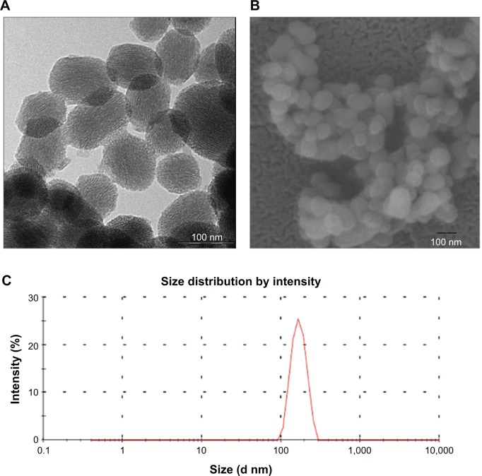
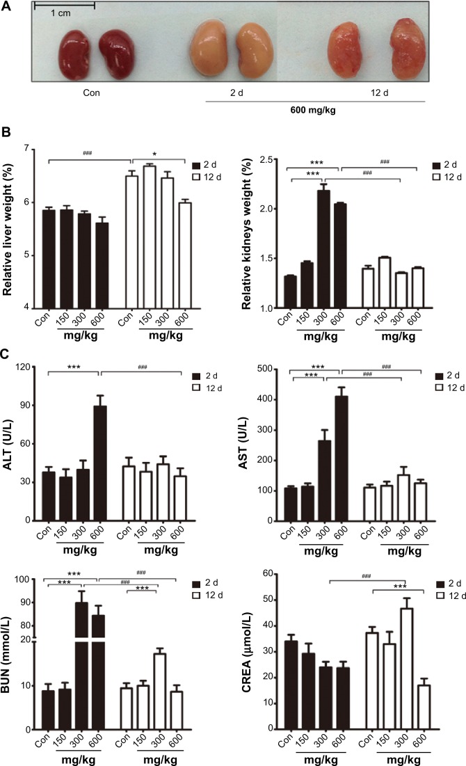
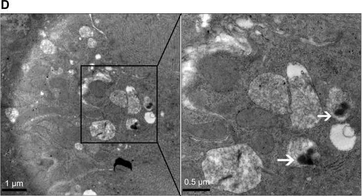
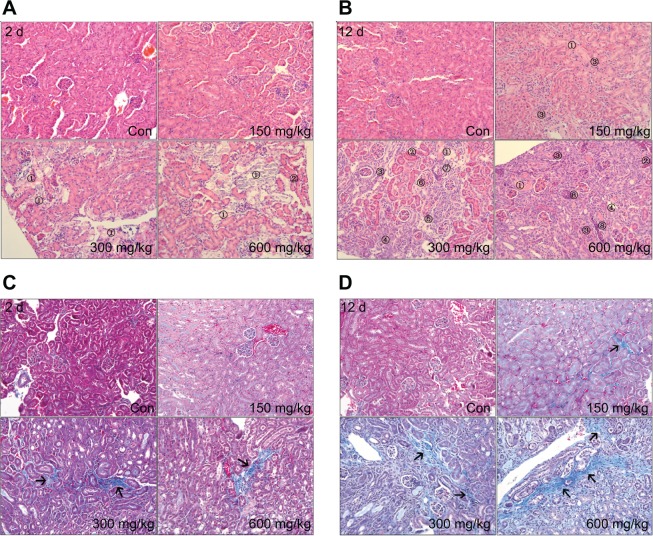
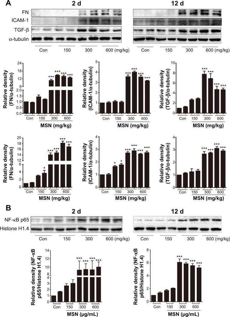
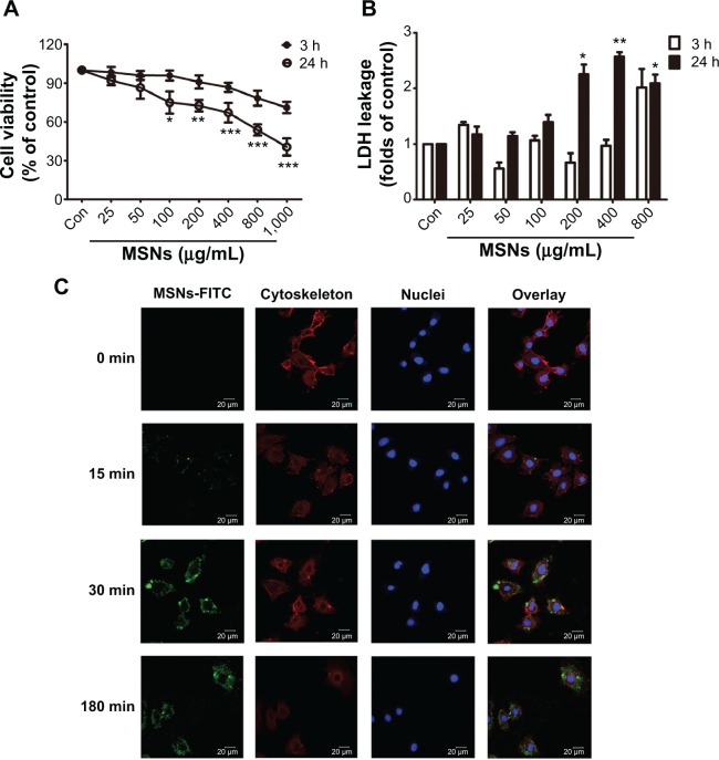
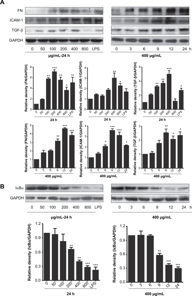
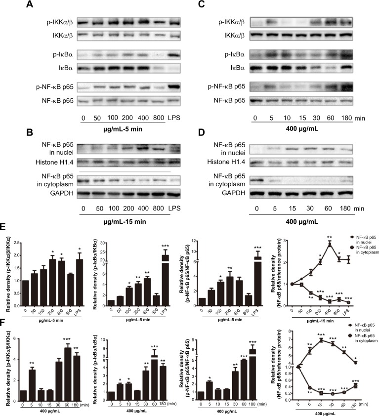
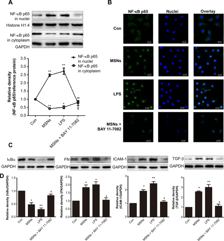
Previous studies have indicated that the nephrotoxicity induced by mesoporous silica nanoparticles (MSNs) is closely related to inflammation. Nuclear factor kappa B (NF-κB), a common rapid transcription factor associated with inflammation, plays an important role in the process of many kidney diseases. Acute toxicity assessment with a high-dose exposure is critical for the development of nanoparticle, as a part of standardized procedures for the evaluation of their toxicity. The present study was undertaken to observe the acute toxicity, predict the potential target organs of MSNs injury, and test the hypothesis that the NF-κB pathway plays a role in mediating the acute kidney injury and renal interstitial fibrosis in mice induced by MSNs. Balb/c mice were intraperitoneally injected with MSNs at concentrations of 150, 300, or 600 mg/kg. All of the animals were euthanized 2 and 12 days after exposure, and the blood and kidney tissues were collected for further studies. In vitro, the cytotoxicity, fibrosis markers, and NF-κB pathway were measured in a normal rat kidney cell line (NRK-52E). Acute kidney injury was induced by MSNs in mice after 2 days, some renal tubules regenerated and renal interstitial fibrosis was also observed. The expression of fibrosis markers and the nuclear translocation of NF-κB p65 in the kidney homogenates increased after exposure to MSNs. The in vitro study showed that MSNs cause cytotoxicity in NRK-52E cells and increased the expression of fibrosis markers. In addition, the NF-κB pathway could be induced, and inhibition of the NF-κB pathway could alleviate the fibrosis caused by MSNs. We conclude that inflammation is a major effector of the acute kidney toxicity induced by MSNs and results in renal interstitial fibrosis, which is mediated by the NF-κB signaling pathway.

Keywords: NF-κB; acute kidney injury; mesoporous silica nanoparticles (MSNs); renal interstitial fibrosis.


Figures

Figure 1
Figure 1
Characterization of MSNs. Notes: Size distribution of MSNs following dispersion in 5% glucose. Particle size distribution evaluated from the corresponding SEM (A) and TEM micrographs (B). Mean particle size (C) of the ordered mesoporous silica. The scale is indicated at the right lower corner. The average diameter of the MSNs was found to be 198.2±21.8 nm in 5% glucose solution, with the particle dispersion index of 0.146±0.067. Abbreviations: MSNs, mesoporous silica nanoparticles; SEM, scanning electron microscopy; TEM, transmission electron micrographs.
Figure 2
Figure 2
Effects of MSNs in Balb/c mice. Notes: The kidney appearances of the treated mice: con, 600 mg/kg (for 2 days) and 600 mg/kg (for 12 days) (A). All of the mice were treated with a single intraperitoneal injection. Relative liver and kidney weights of the treated mice (B). Biochemical analysis of aspartate aminotransferase (AST), alanine aminotransferase (ALT), blood urea nitrogen (BUN), and creatinine (CREA) (C). The TEM photos of the renal tubular epithelial cell after treatment with 300 mg/kg MSNs for 2 days were enlarged 23,000- and 46,000-fold; MSNs indicated by arrows (D). The data are expressed as the means ± standard error of the mean (n=5). *P<0.05 and ***P<0.001 versus intragroup con; #P<0.05 and ###P<0.001 among groups. Abbreviations: MSNs, mesoporous silica nanoparticles; SEM, scanning electron microscopy; TEM, transmission electron micrographs; d, day(s); con, control.
Figure 2
Figure 2
Effects of MSNs in Balb/c mice. Notes: The kidney appearances of the treated mice: con, 600 mg/kg (for 2 days) and 600 mg/kg (for 12 days) (A). All of the mice were treated with a single intraperitoneal injection. Relative liver and kidney weights of the treated mice (B). Biochemical analysis of aspartate aminotransferase (AST), alanine aminotransferase (ALT), blood urea nitrogen (BUN), and creatinine (CREA) (C). The TEM photos of the renal tubular epithelial cell after treatment with 300 mg/kg MSNs for 2 days were enlarged 23,000- and 46,000-fold; MSNs indicated by arrows (D). The data are expressed as the means ± standard error of the mean (n=5). *P<0.05 and ***P<0.001 versus intragroup con; #P<0.05 and ###P<0.001 among groups. Abbreviations: MSNs, mesoporous silica nanoparticles; SEM, scanning electron microscopy; TEM, transmission electron micrographs; d, day(s); con, control.
Figure 3
Figure 3
Pathologic changes induced by MSNs in Balb/c mice. Notes: Kidney injury and renal interstitial fibrosis in the control and MSN-treated groups after 2 days and 12 days were detected by hematoxylin and eosin (A and B) and Masson staining (C and D) (magnification 200×), respectively. Because various lesions were observed, numbers were used to indicate the local lesions. 1) Tubular necrosis, 2) tubular eosinophil degeneration, 3) renal interstitial fibrosis, 4) tubular regeneration, 5) renal interstitial lymphocytic infiltration, 6) mesangial cells proliferation, 7) glomerular basement membrane thickening, 8) cellular casts in tubules. In (C and D), the arrows indicate the collagen fiber accumulation in the kidney, which was stained blue with Masson staining. Abbreviations: MSNs, mesoporous silica nanoparticles; d, day(s); con, control.
Figure 4
Figure 4
MSNs increased kidney fibrosis in Balb/c mice 2 days and 12 days after injection. Notes: The proteins (20 εg) of the total lysates extracted from the left kidney were subjected to SDS-PAGE followed by Western blot with the indicated antibodies (A and B). α-Tubulin was used as the loading control. The blots were then quantified by densitometry. The data are expressed as the means ± standard error of the mean. The results are representative of three independent experiments. (*P<0.05 and ***P<0.001 versus con intragroup, ANOVA). Abbreviations: MSNs, mesoporous silica nanoparticles; d, day(s); ANOVA, analysis of variance; NF-κB, nuclear factor kappaa B; con, control.
Figure 5
Figure 5
Effects of MSNs on NRK-52E cells. Notes: (A) NRK-52E cells were treated with MSNs at various concentrations for 3 h and 24 h. The cell viability was determined by an MTT assay. The viability of the cells that were not treated with MSNs is defined as 100%. (B) For the LDH release assay, NRK-52E cells were treated as in (A), and the conditional medium was harvested for LDH assay. The data are expressed as the means ± standard error of the mean. The results are representative of three independent experiments. (*P<0.05, **P<0.01, and ***P<0.001 versus con, ANOVA). (C) The uptake of MSNs by NRK-52E cells was detected at 0, 15, 30, and 180 min. MSNs were stained green due to their labeling with FITC, the cytoskeleton presented red staining due to Phalloidin-TRITC, and the nuclei were stained blue due to DAPI staining. Abbreviations: MSNs, mesoporous silica nanoparticles; NRK, normal rat kidney; h, hour(s); min, minutes; LDH, lactate dehydrogenase; ANOVA, analysis of variance; FITC, fluorescein isothiocyanate; TRITC, tetramethylrhodamine; DAPI, 4′,6-diamidino-2-phenylindole; con, control.
Figure 6
Figure 6
Fibrosis and inflammation induced by MSNs in NRK-52E cells. Notes: Fibrosis (A) and inflammation (B) induced by MSNs in NRK-52E cells. NRK-52E cells were treated with the indicated concentration of MSNs for 24 h and treated with 400 εg/mL MSNs for the indicated times. The proteins (20 εg) of the total lysates extracted from the NRK-52E cells were subjected to SDS-PAGE followed by Western blot with the indicated antibodies. GAPDH was used as the loading control. The blots were then quantified by densitometry. The data are expressed as the means ± standard error of the mean. The results are representative of three independent experiments. (*P<0.05, **P<0.01, and ***P<0.001 versus con, ANOVA). Abbreviations: MSNs, mesoporous silica nanoparticles; NRK, normal rat kidney; h, hour(s); ANOVA, analysis of variance; con, control; LPS, Lipopolysaccharides.
Figure 7
Figure 7
Effects of MSNs on the activation of the NF-κB signaling pathway. Notes: NRK-52E cells were treated with the indicated concentrations of MSNs for 5 min (A),15 min (B), and 30 min (E); or treated with 400 εg/mL MSNs for the indicated time (C, D and F). The proteins (20 εg) of the total lysates and the nuclear (15 εg) and cytoplasm proteins (15 εg) extracted from NRK-52E cells were subjected to 10% SDS-PAGE followed by Western blot with the indicated antibodies. NRK-52E cells treated with 2 εg/mL LPS served as the positive control. Histone H1.4 and GAPDH were used as the loading controls for the nuclear and cytoplasmic proteins, respectively. The blots were then quantified by densitometry. TNF-α, IL-1β, and IL-6 production was assessed using ELISA assay (E and F). The data are presented as the means ± standard error of the mean from three independent experiments. (*P<0.05, **P<0.01, and ***P<0.001 versus con, ANOVA). Abbreviations: MSNs, mesoporous silica nanoparticles; NF-κB, nuclear factor kappa B; NRK, normal rat kidney; min, minutes; ANOVA, analysis of variance; ELISA, enzyme-linked immunosorbent assay; con, control; LPS, Lipopolysaccharides.
Figure 8
Figure 8
Reduction of the fibrosis by MSNs on NRK-52E cells through inhibition of the NF-κB pathway. Notes: The changes in the protein expression (A) and immunofluorescence staining (B) of NF-κB p65 in the nuclei and cytoplasm were evaluated through Western blot analysis and confocal microscopy. MSNs: 400 εg/mL MSNs for 30 min; LPS: 2 εg/mL LPS for 30 min; MSNs + BAY 11-7082: pre-treatment with 3 εM BAY 11-7082 for 1 h followed by treatment with MSNs for 30 min. The fibrosis reduction was measured by Western blot with the indicated antibodies (C). MSNs: 400 εg/mL MSNs for 24 h; LPS: 2 εg/mL LPS for 24 h; MSNs + BAY 11-7082: pre-treatment with 3 εM BAY 11-7082 for 2 h followed by treatment with MSNs for 24 h. Histone H1.4 and GAPDH were used as the loading control for the nuclear and cytoplasmic proteins, respectively. TNF-α, IL-1β, and IL-6 production (D) was assessed using ELISA assay. MSNs: 400 εg/mL MSNs for 2 h; LPS: 2 εg/mL LPS for 30 min; MSNs + BAY 11-7082: pre-treatment with 3 εM BAY 11-7082 for 1 h followed by treatment with MSNs for 2 h. The data are expressed as the means ± standard error of the mean. The results are representative of three independent experiments. (*P<0.05, and **P<0.01 versus con, #P<0.05, and ##P<0.01 versus MSNs, ANOVA). Abbreviations: MSNs, mesoporous silica nanoparticles; NF-κB, nuclear factor kappa B; NRK, normal rat kidney; min, minutes; h, hour(s); ELISA, enzyme-linked immunosorbent assay; ANOVA, analysis of variance; con, control; LPS, Lipopolysaccharides.

References

    1. Vallhov H, Gabrielsson S, Strømme M, Scheynius A, Garcia-Bennett AE. Mesoporous silica particles induce size dependent effects on human dendritic cells. Nano Lett. 2007;7(12):3576–3582. - PubMed
    1. Chung TH, Wu SH, Yao M, et al. The effect of surface charge on the uptake and biological function of mesoporous silica nanoparticles in 3T3-L1 cells and human mesenchymal stem cells. Biomaterials. 2007;28(19):2959–2966. - PubMed
    1. Carino IS, Pasqua L, Testa F, et al. Silica-based mesoporous materials as drug delivery system for methotrexate release. Drug Deliv. 2007;14(8):491–495. - PubMed
    1. Gu J, Fan W, Shimojima A, Okubo T. Organic-inorganic mesoporous nanocarriers integrated with biogenic ligands. Small. 2007;3(10):1740–1744. - PubMed
    1. Yun HS, Park JW, Kim SH, Kim YJ, Jang JH. Effect of the pore structure of bioactive glass balls on biocompatibility in vitro and in vivo. Acta Biomater. 2011;7(6):2651–2660. - PubMed

Publication types

MeSH terms