Review
. 2014 Dec 16:8:404.
doi: 10.3389/fnins.2014.00404. eCollection 2014.

The rights and wrongs of blood-brain barrier permeability studies: a walk through 100 years of history

Affiliations
Review

The rights and wrongs of blood-brain barrier permeability studies: a walk through 100 years of history

Norman R Saunders et al. Front Neurosci. .

Abstract

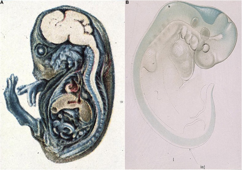
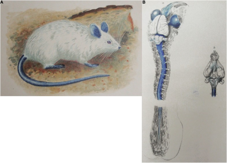
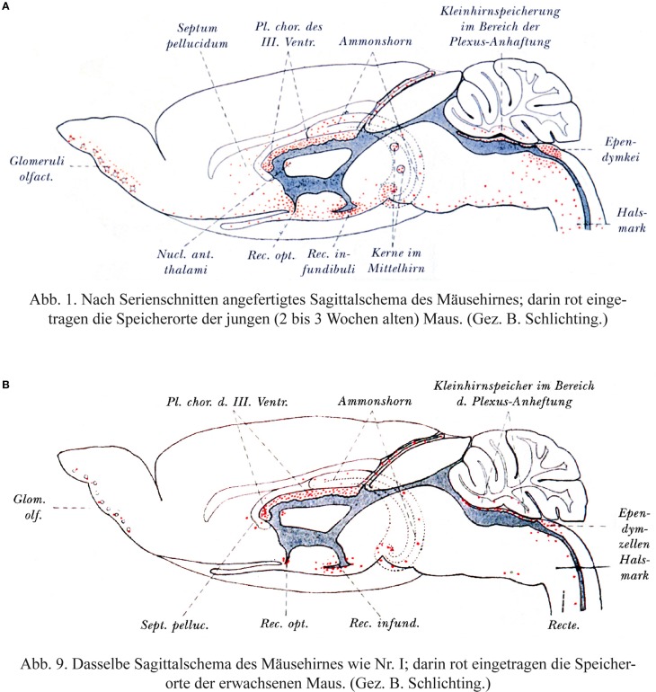
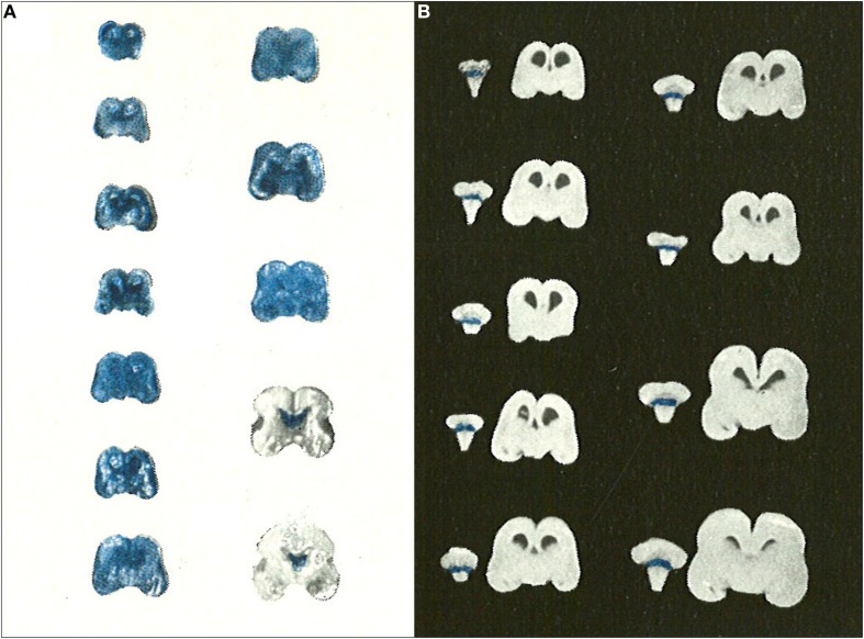
Careful examination of relevant literature shows that many of the most cherished concepts of the blood-brain barrier are incorrect. These include an almost mythological belief in its immaturity that is unfortunately often equated with absence or at least leakiness in the embryo and fetus. The original concept of a blood-brain barrier is often attributed to Ehrlich; however, he did not accept that permeability of cerebral vessels was different from other organs. Goldmann is often credited with the first experiments showing dye (trypan blue) exclusion from the brain when injected systemically, but not when injected directly into it. Rarely cited are earlier experiments of Bouffard and of Franke who showed methylene blue and trypan red stained all tissues except the brain. The term "blood-brain barrier" "Blut-Hirnschranke" is often attributed to Lewandowsky, but it does not appear in his papers. The first person to use this term seems to be Stern in the early 1920s. Studies in embryos by Stern and colleagues, Weed and Wislocki showed results similar to those in adult animals. These were well-conducted experiments made a century ago, thus the persistence of a belief in barrier immaturity is puzzling. As discussed in this review, evidence for this belief, is of poor experimental quality, often misinterpreted and often not properly cited. The functional state of blood-brain barrier mechanisms in the fetus is an important biological phenomenon with implications for normal brain development. It is also important for clinicians to have proper evidence on which to advise pregnant women who may need to take medications for serious medical conditions. Beliefs in immaturity of the blood-brain barrier have held the field back for decades. Their history illustrates the importance of taking account of all the evidence and assessing its quality, rather than selecting papers that supports a preconceived notion or intuitive belief. This review attempts to right the wrongs. Based on careful translation of original papers, some published a century ago, as well as providing discussion of studies claiming to show barrier immaturity, we hope that readers will have evidence on which to base their own conclusions.

Keywords: blood-brain barrier; blood-cerebrospinal fluid barrier; embryo; fetus; newborn; permeability; tight junctions; transporters.


Figures

Figure 1
Figure 1
Early demonstration of blood-brain barrier phenomenon in developing brain. (A) Mid gestation guinea pig embryo injected with trypan blue (Wislocki, 1920). Note lack of staining of brain and spinal cord as previously described in adult animals injected with trypan blue (Goldmann, 1909), methylene blue (Bouffard, 1906) and trypan red (Franke, 1905). (B) Very early pig embryo (9 mm, E19) injected with isotonic sodium ferrocyanide into spinal canal (arrow) treated with acid (Prussian blue reaction). Note that blue reaction product is confined to CSF, with a small amount of diffusion into brain stem and mid brain tissue, but no staining of somatic tissue. This indicates that the CNS is already a closed compartment separate from the rest of the embryo. From Weed (1917b).
Figure 2
Figure 2
Goldmann's trypan blue injection experiments. (A) Adult rat following systemic injection of trypan blue solution (Goldmann's “1st experiment”). From Goldmann (1909). (B) Brain and spinal cord of adult rat following lumbar subarachnoid injection of 0.5 ml, 0.5% trypan blue (Goldmann's “2nd experiment”). From Goldmann (1913).
Figure 3
Figure 3
Behnsen's much cited trypan blue experiments. (A) Brain from postnatal mouse injected systemically 3 times with trypan blue at P4–P14. (B) Adult mouse brain following systemic injection of trypan blue. Note that as described by Behnsen the dye staining in the postnatal brain is in the same regions as in the adult brain, but more extensive. This is probably a visual artifact due to the use of an outline of an adult mouse brain for both ages. In postnatal mouse brain (P14) the cerebral cortex and cerebellum are less developed than illustrated here. Note also that in the postnatal brain there does not appear to be any dye staining of the neocortex-the region of the developing brain with the least mature blood vessels. From Behnsen (1927).
Figure 4
Figure 4
Gröntoft's dye experiments with postmortem aborted human fetuses. (A) Human fetuses (14 cm-18 weeks to 30 cm-31 weeks gestation) injected with trypan blue solution 30 min after caesarian section delivery. (B) Human fetuses (14 cm-18 weeks to 30 cm-31 weeks gestation) injected with trypan blue solution 10 min after caesarian section delivery. Note that for the shorter period of anoxia none of the brains stained with trypan blue indicating intact blood-brain barrier to trypan blue even in immature human fetuses. Reproduced with permission from Gröntoft (1954), © Wiley.
Figure 5
Figure 5
Kernicterus in brain of neonatal monkey with jaundice. Note staining of putamen (one of the basal ganglia), which does not occur in adult brain as jaundice in adults, is generally due to high levels of conjugated bilirubin to which the blood brain barrier is impermeable. From Windle (1969).

References

    1. Abbott N. J., Rönnbäck L., Hansson E. (2006). Astrocyte-endothelial interactions at the blood-brain barrier. Nat. Rev. Neurosci. 7, 41–53. 10.1038/nrn1824 - DOI - PubMed
    1. Adinolfi M. (1985). The development of the human blood-brain barrier. Dev. Med. Child. Neurol. 27, 532–537. 10.1111/j.1469-8749.1985.tb04581.x - DOI - PubMed
    1. Adinolfi M., Beck S. E., Haddad S. A., Seller M. J. (1976). Permeability of the blood-cerebrospinal fluid barrier to plasma proteins during foetal and perinatal life. Nature 259, 140–141. 10.1038/259140a0 - DOI - PubMed
    1. Adinolfi M., Haddad S. A. (1977). Levels of plasma proteins in human and rat fetal CSF and the development of the blood-CSF barrier. Neuropädiatrie 8, 345–353. 10.1055/s-0028-1091530 - DOI - PubMed
    1. Ahlfors C. E., Wennberg R. P. (2004). Bilirubin-albumin binding and neonatal jaundice. Semin. Perinatol. 28, 334–339. 10.1053/j.semperi.2004.09.002 - DOI - PubMed