. 2014 Dec 17:8:428.
doi: 10.3389/fncel.2014.00428. eCollection 2014.

Glutamic acid decarboxylase 67 expression by a distinct population of mouse vestibular supporting cells

Affiliations

Glutamic acid decarboxylase 67 expression by a distinct population of mouse vestibular supporting cells

Elisa Tavazzani et al. Front Cell Neurosci. .

Erratum in

Abstract

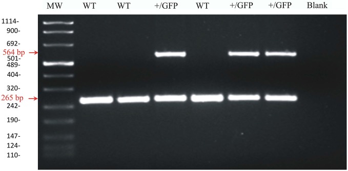
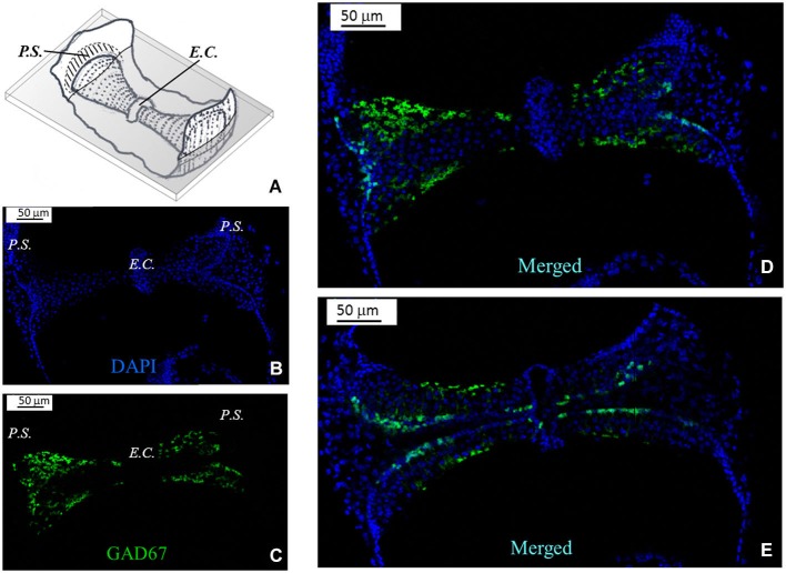
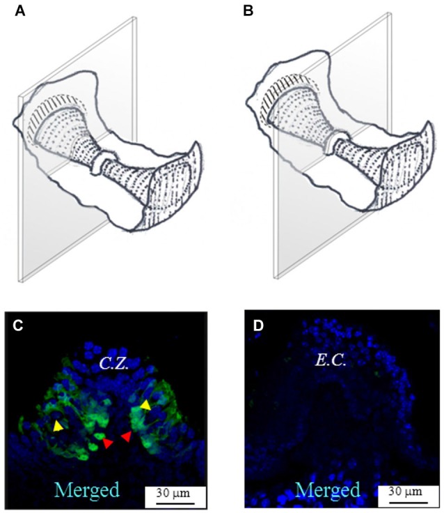
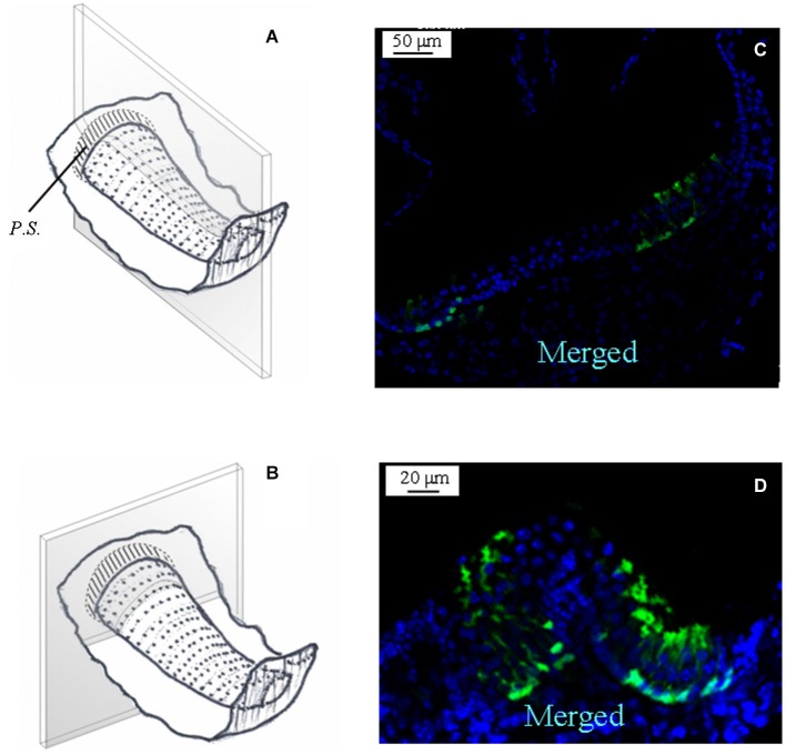
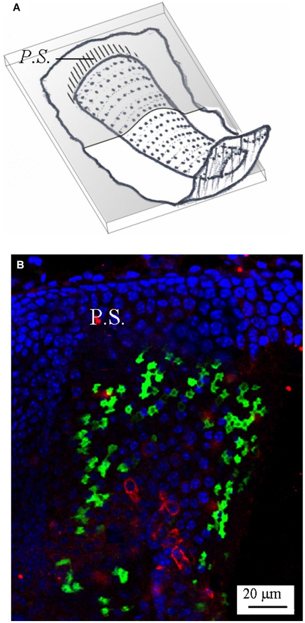
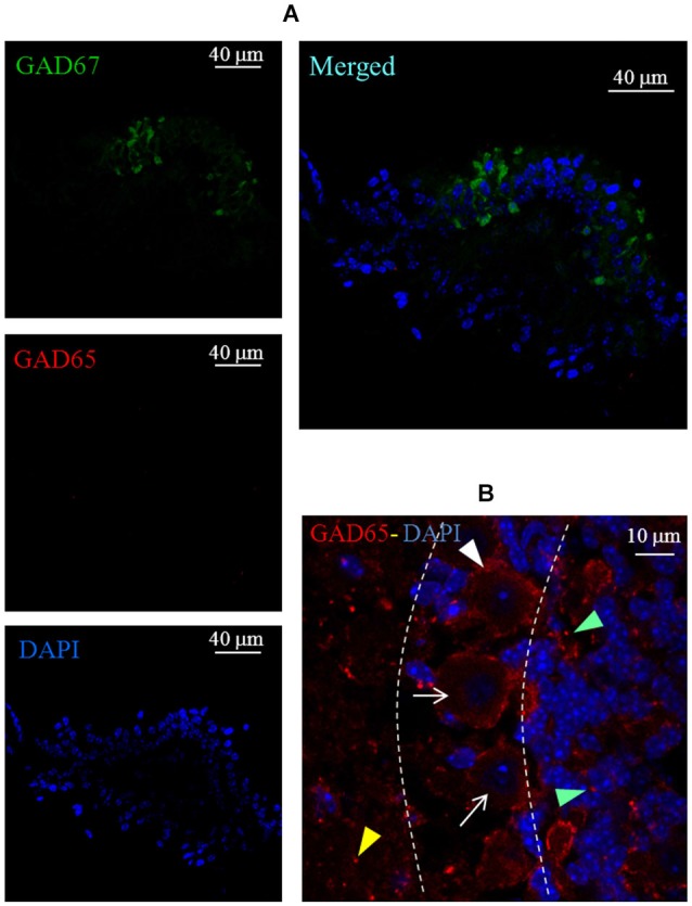
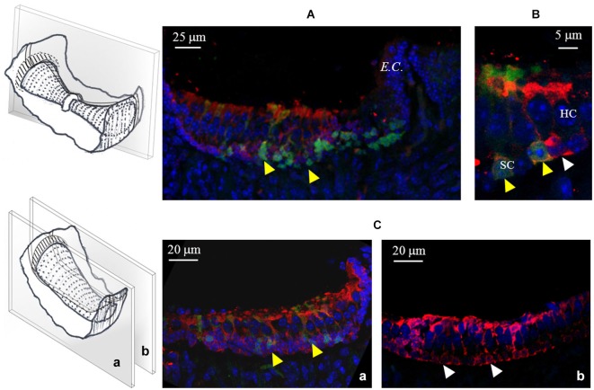
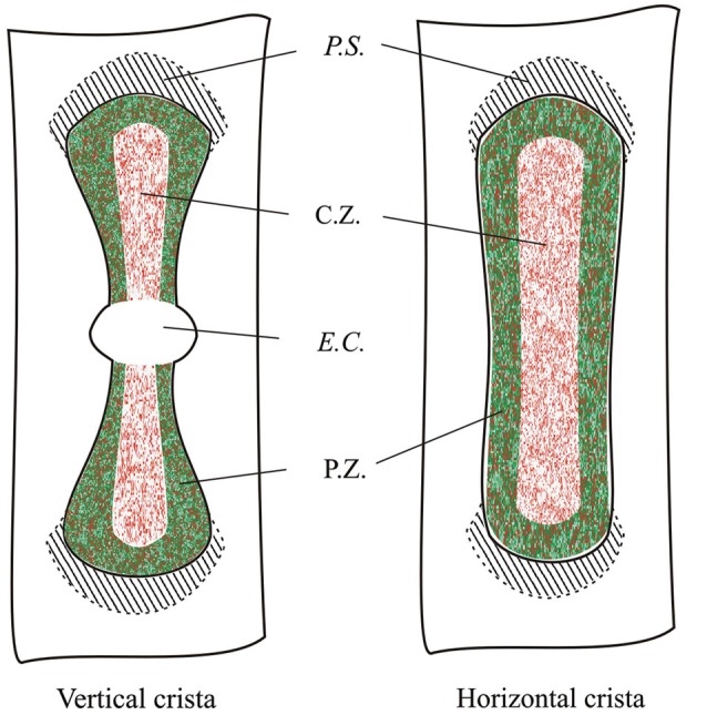
The function of the enzyme glutamate decarboxylase (GAD) is to convert glutamate in γ-aminobutyric acid (GABA). Glutamate decarboxylase exists as two major isoforms, termed GAD65 and GAD67, that are usually expressed in GABA-containing neurons in the central nervous system. GAD65 has been proposed to be associated with GABA exocytosis whereas GAD67 with GABA metabolism. In the present immunofluorescence study, we have investigated the presence of the two GAD isoforms in the semicircular canal cristae of wild type and GAD67-GFP knock-in mice. While no evidence for GAD65 expression was found, GAD67 was detected in a distinct population of peripherally-located supporting cells, but not in hair cells or in centrally-located supporting cells. GABA, on the other hand, was found in all supporting cells. The present result indicate that only a discrete population of supporting cells use GAD67 to synthesize GABA. This is the first report of a marker that allows to distinguish two populations of supporting cells in the vestibular epithelium. On the other hand, the lack of GABA and GAD enzymes in hair cells excludes its involvement in afferent transmission.

Keywords: GAD67-GFP; crista ampullaris; hair cells; supporting cells; vestibular.


Figures

Figure 1
Figure 1
Agarose gel image of PCR products for GAD67 and GAD67-GFP from wild-type and heterozygous GAD67-GFP knock-in mice. Representative PCR products for GAD67 (265 pb) and GAD67-GFP (564 pb) were observed in +/GFP lanes (n = 3) while only the PCR product for GAD67 was present in the wild type lanes (WT, n = 3). The control lane (blank) was negative as expected. The left lane (MW) shows DNA molecular weight markers.
Figure 2
Figure 2
GAD67 expression in the mouse vertical crista. (A) Schematic representation showing a representative slice (gray) of the crista ampullaris. Dots represent hair bundles protruding from the crista surface. E.C.: eminentia cruciata; P.S.: planum semilunatum. (B,C,D) Photomicrographs of a superficial confocal section of the slice schematized in (A) showing the cell nuclei (blue; DAPI), the cellular expression of GAD67 (green; GFP), and the merged image, respectively. Light blue indicates co-localization of nuclei and GFP, which seems poor because GAD67 is expressed in the cytoplasm. Most cell bodies at this confocal level appear sectioned transversally. (E) Photomicrograph of a lower confocal section of the same specimen (merged image). Most cell bodies appear now sectioned longitudinally, as inferred from their elongated shape.
Figure 3
Figure 3
GAD67 expression in transverse slices of the mouse vertical crista. (A,B) Schematic representations showing the plane of the transverse sections. (C,D) Photomicrographs of the confocal sections showing the expression of GAD67. C.Z.: central zone. Note the absence of GAD67 at the most central zone in (C), and the complete absence of GAD67 in the E.C. in (D). The red arrowheads indicate the position of supporting cell nuclei, while the yellow arrowheads the position of hair cell nuclei.
Figure 4
Figure 4
GAD67 expression in longitudinal slices of the mouse vertical crista. (A) Schematic representation showing the plane of the slice. Note that the section is not through the center of the crista (i.e., it is not a medial section). (B) Merged photomicrograph showing the expression of GAD67. Note the absence of GAD67 expression in the E.C. (white arrow). (C) Enlargement of a portion of the same image as in (B), showing in better detail the shape and position of the cells expressing GAD67. The white arrow points at the nuclear region of a supporting cell. The red arrow points at a nucleus of a hair cell.
Figure 5
Figure 5
GAD67 expression in the mouse horizontal crista. (A,B) Schematic representations showing the plane of the longitudinal and transverse sections shown in (C) and (D). Note the absence, also in the horizontal crista, of GAD67 expression in the central zone of the sensory epithelium.
Figure 6
Figure 6
Calbindin expression in the mouse horizontal crista. (A) Schematic representation showing the plane of the section. (B) Photomicrograph of a longitudinal confocal section showing the cell nuclei (blue; DAPI), GAD67 (green; GFP), and calbindin (red; antibody) expression—merged image. Note that most calbindin immunolabeling is concentrated in the central zone, where it marks several calyx endings.
Figure 7
Figure 7
Lack of GAD65 expression in the mouse crista. (A) Photomicrographs of a crista transverse confocal section showing the expression of GAD67 (green), GAD65 (red), cell nuclei (blue; DAPI), and the merged image. Note the absence of GAD65 immunofluorescence. (B) Photomicrograph of a confocal section of a mouse cerebellum slice taken as positive control for GAD65 antibody labeling (red). Note the weak and diffuse immunofluorescence for GAD65 inside Purkinje cells (arrows) and immunofluorescent spots labeling around Purkinje cells (white arrowhead), in the molecular layer (yellow arrowhead) and in the granular layer (green arrowheads). Dashed lines indicate the bounders among the three layers.
Figure 8
Figure 8
GABA expression in the mouse cristae. (A) Photomicrograph of a confocal section from a longitudinal slice of a vertical crista. As shown in the schematic representation on the left, the picture refers to the peripheral zone. Note the co-localization (yellow arrowheads) of GABA (red) and GAD67 (green) in many supporting cells. (B) Higher magnification from a different vertical crista section showing GABA and GAD67 co-localization (yellow arrowheads) or GABA-only expression (white arrowhead) in three different supporting cells. (C) Confocal images from two longitudinal slices of a horizontal crista depicting the peripheral (a) and the central (b) zone. Most peripheral supporting cells co-express GABA and GAD67, while central supporting cells only express GABA. No GABA expression was ever found in hair cells.
Figure 9
Figure 9
Topographical distribution of GAD67 and GABA in the mouse vertical and horizontal cristae. Schematic representation showing the expression of GAD67 (green) by peripheral supporting cells as inferred by the confocal experiments. Red dots indicate supporting cell expressing GABA. C.Z.: central zone; P.Z.: peripheral zone; P.S.: planum semilunatum; E.C.: eminentia cruciata.

References

    1. Bonsacquet J., Brugeaud A., Compan V., Desmadryl G., Chabbert C. (2006). AMPA type glutamate receptor mediates neurotransmission at turtle vestibular calyx synapse. J. Physiol. 576, 63–71. 10.1113/jphysiol.2006.116467 - DOI - PMC - PubMed
    1. Bu D. F., Erlander M. G., Hitz B. C., Tillakaratne N. J., Kaufman D. L., Wagner-McPherson C. B., et al. . (1992). Two human glutamate decarboxylases, 65-kDa GAD and 67-kDa GAD, are each encoded by a single gene. Proc. Natl. Acad. Sci. U S A 89, 2115–2119. 10.1073/pnas.89.6.2115 - DOI - PMC - PubMed
    1. Dalet A., Bonsacquet J., Gaboyard-Niay S., Calin-Jageman I., Chidavaenzi R. L., Venteo S., et al. . (2012). Glutamate transporters EAAT4 and EAAT5 are expressed in vestibular hair cells and calyx endings. PLoS One 7:e46261. 10.1371/journal.pone.0046261 - DOI - PMC - PubMed
    1. Desai S. S., Ali H., Lysakowski A. (2005). Comparative morphology of rodent vestibular periphery. II. Cristae ampullares. J. Neurophysiol. 93, 267–280. 10.1152/jn.00747.2003 - DOI - PubMed
    1. Eatock R. A., Songer J. E. (2011). Vestibular hair cells and afferents: two channels for head motion signals. Annu. Rev. Neurosci. 34, 501–534. 10.1146/annurev-neuro-061010-113710 - DOI - PubMed