

Satellite cell activity is differentially affected by contraction mode in human muscle following a work-matched bout of exercise
- PMID: 25566087
- PMCID: PMC4263080
- DOI: 10.3389/fphys.2014.00485
Satellite cell activity is differentially affected by contraction mode in human muscle following a work-matched bout of exercise
Abstract
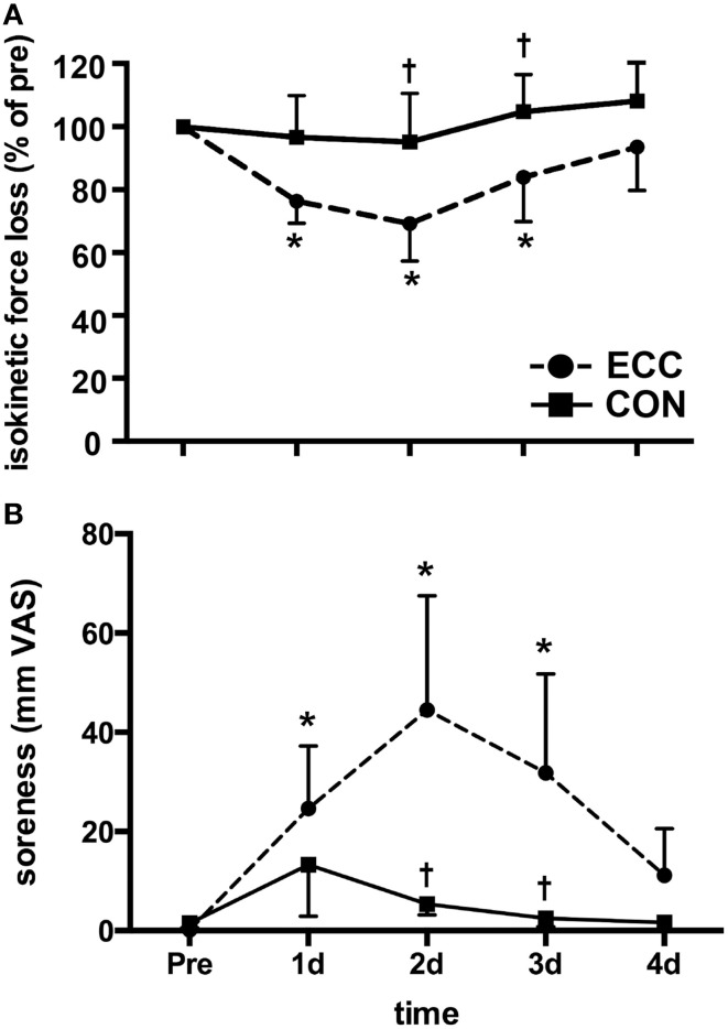
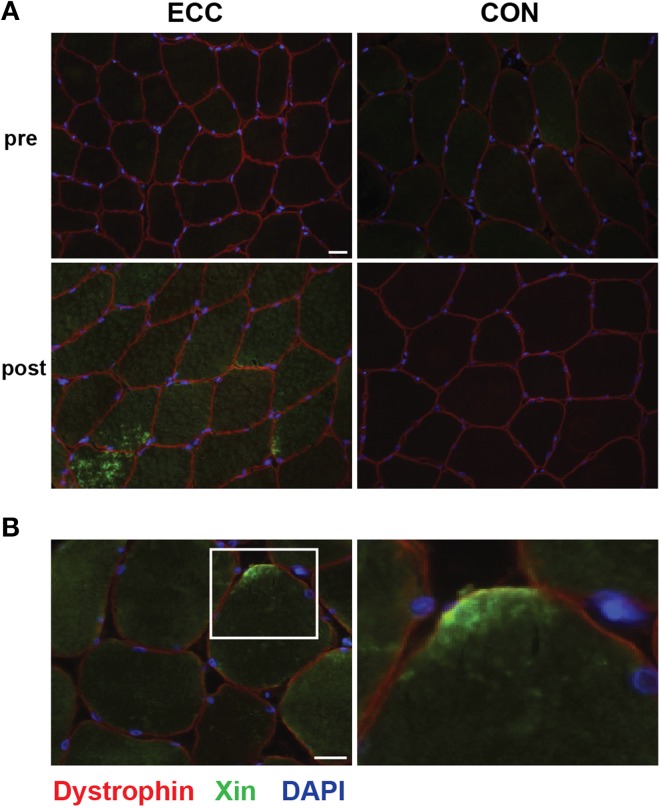
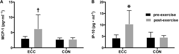
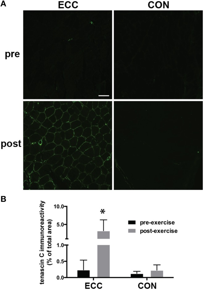
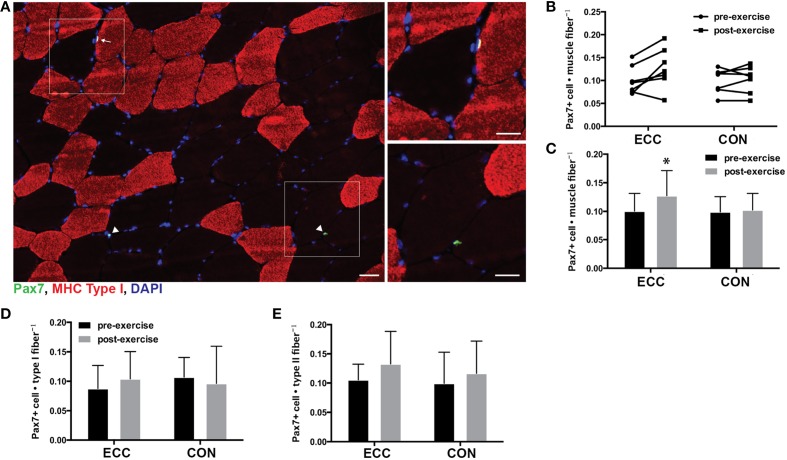
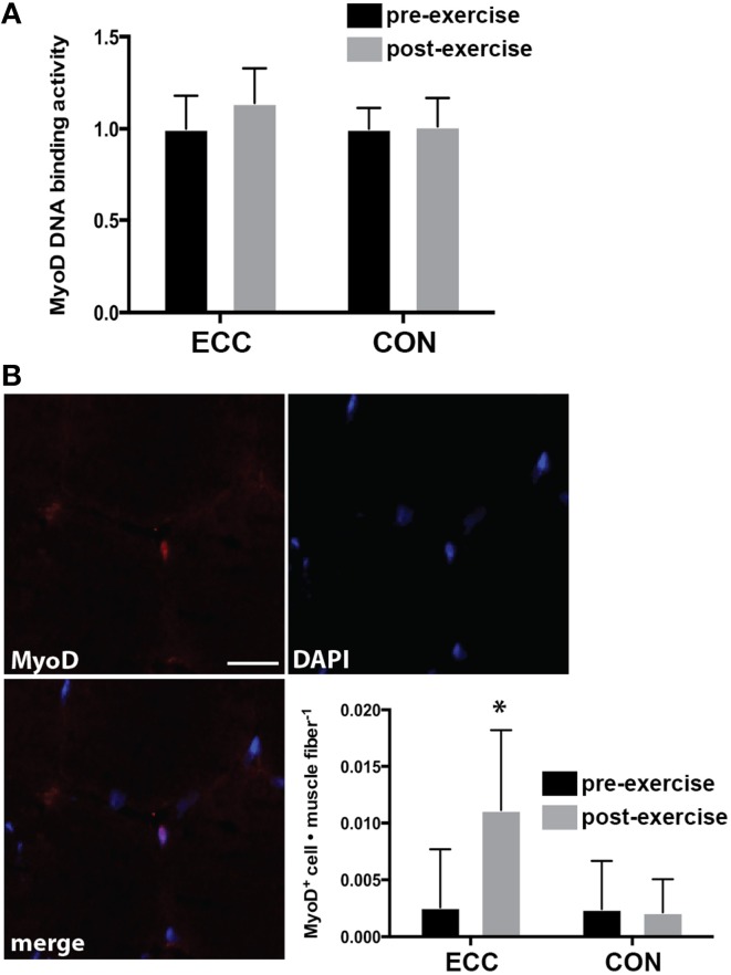
Optimal repair and adaptation of skeletal muscle is facilitated by resident stem cells (satellite cells). To understand how different exercise modes influence satellite cell dynamics, we measured satellite cell activity in conjunction with markers of muscle damage and inflammation in human skeletal muscle following a single work- and intensity-matched bout of eccentric (ECC) or concentric contractions (CON). Participants completed a single bout of ECC (n = 7) or CON (n = 7) of the knee extensors. A muscle biopsy was obtained before and 24 h after exercise. Functional measures and immunohistochemical analyses were used to determine the extent of muscle damage and indices of satellite cell activity. Cytokine concentrations were measured using a multiplexed magnetic bead assay. Isokinetic peak torque decreased following ECC (p < 0.05) but not CON. Greater histological staining of the damage marker Xin was observed in muscle samples of ECC vs. CON. Tenasin C immunoreactivity increased 15 fold (p < 0.01) following ECC and was unchanged following CON. The inflammatory cytokines interferon gamma-induced protein 10 (IP-10) and monocyte chemotactic protein 1 (MCP-1) increased pre- to post-ECC (4.26 ± 1.4 vs. 10.49 ± 5.8 pg/ml, and 3.06 ± 0.7 vs. 6.25 ± 4.6 pg/ml, respectively; p < 0.05). There was no change in any cytokine post-CON. Satellite cell content increased 27% pre- to post-ECC (0.10 ± 0.031 vs. 0.127 ± 0.041, respectively; p < 0.05). There was no change in satellite cell number in CON (0.099 ± 0.027 vs. 0.102 ± 0.029, respectively). There was no fiber type-specific satellite cell response following either exercise mode. ECC but not CON resulted in an increase in MyoD positive nuclei per myofiber pre- to post-exercise (p < 0.05), but there was no change in MyoD DNA binding activity in either condition. In conclusion, ECC but not CON results in functional and histological evidence of muscle damage that is accompanied by increased satellite cell activity 24 h post-exercise.
Keywords: MyoD; Pax7; concentric; eccentric; resistance exercise; stem cell.
Figures
Similar articles
-
Eccentric Overload during Resistance Exercise: A Stimulus for Enhanced Satellite Cell Activation.Med Sci Sports Exerc. 2022 Mar 1;54(3):388-398. doi: 10.1249/MSS.0000000000002818. Med Sci Sports Exerc. 2022. PMID: 34690286
-
Force and EMG signal patterns during repeated bouts of concentric or eccentric muscle actions.Acta Physiol Scand. 1990 Mar;138(3):263-71. doi: 10.1111/j.1748-1716.1990.tb08846.x. Acta Physiol Scand. 1990. PMID: 2327260
-
Strength Training Effects on Muscular Regeneration after ACL Reconstruction.Med Sci Sports Exerc. 2018 Jun;50(6):1152-1161. doi: 10.1249/MSS.0000000000001564. Med Sci Sports Exerc. 2018. PMID: 29389836 Clinical Trial.
-
Skeletal Muscle Remodeling in Response to Eccentric vs. Concentric Loading: Morphological, Molecular, and Metabolic Adaptations.Front Physiol. 2017 Jul 4;8:447. doi: 10.3389/fphys.2017.00447. eCollection 2017. Front Physiol. 2017. PMID: 28725197 Free PMC article. Review.
-
Eccentric Training Improves Body Composition by Inducing Mechanical and Metabolic Adaptations: A Promising Approach for Overweight and Obese Individuals.Front Physiol. 2018 Aug 7;9:1013. doi: 10.3389/fphys.2018.01013. eCollection 2018. Front Physiol. 2018. PMID: 30131705 Free PMC article. Review.
Cited by
-
Molecular clocks, satellite cells, and skeletal muscle regeneration.Am J Physiol Cell Physiol. 2023 Jun 1;324(6):C1332-C1340. doi: 10.1152/ajpcell.00073.2023. Epub 2023 May 15. Am J Physiol Cell Physiol. 2023. PMID: 37184229 Free PMC article. Review.
-
Restoring the regenerative balance in neuromuscular disorders: satellite cell activation as therapeutic target in Pompe disease.Ann Transl Med. 2019 Jul;7(13):280. doi: 10.21037/atm.2019.04.48. Ann Transl Med. 2019. PMID: 31392192 Free PMC article. Review.
-
Preconditioning Contractions Suppress Muscle Pain Markers after Damaging Eccentric Contractions.Pain Res Manag. 2018 Oct 14;2018:3080715. doi: 10.1155/2018/3080715. eCollection 2018. Pain Res Manag. 2018. PMID: 30405861 Free PMC article.
-
Cycle training modulates satellite cell and transcriptional responses to a bout of resistance exercise.Physiol Rep. 2016 Sep;4(18):e12973. doi: 10.14814/phy2.12973. Physiol Rep. 2016. PMID: 27650251 Free PMC article.
-
Unraveling the bidirectional relationship between muscle inflammation and satellite cells activity: influencing factors and insights.J Muscle Res Cell Motil. 2025 Mar;46(1):35-51. doi: 10.1007/s10974-024-09683-7. Epub 2024 Nov 7. J Muscle Res Cell Motil. 2025. PMID: 39508952 Review.
References
LinkOut - more resources
Full Text Sources
Other Literature Sources
Research Materials
Miscellaneous