Review
. 2014 Dec 17:5:643.
doi: 10.3389/fimmu.2014.00643. eCollection 2014.

Monitoring Circulating γδ T Cells in Cancer Patients to Optimize γδ T Cell-Based Immunotherapy

Affiliations
Review

Monitoring Circulating γδ T Cells in Cancer Patients to Optimize γδ T Cell-Based Immunotherapy

Hans-Heinrich Oberg et al. Front Immunol. .

Abstract

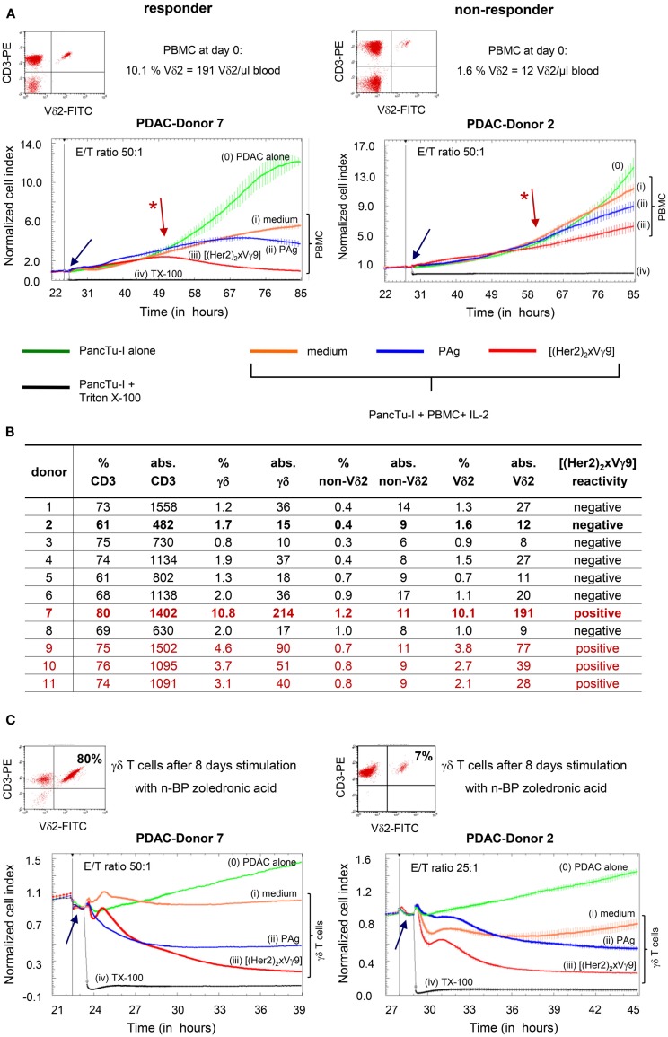
The success of γδ T cell-based immunotherapy, where the cytotoxic activity of circulating γδ T lymphocytes is activated by nitrogen-containing bisphosphonates (n-BP), or possibly by bispecific antibodies or the combination of both, requires a profound knowledge of patients' γδ T cells. A possible influence of radio- or chemotherapy on γδ T cells as well as their reported exhaustion after repetitive treatment with n-BP or their lack of response to various cancers can be easily determined by the monitoring assays described in this perspective article. Monitoring the absolute cell numbers of circulating γδ T cell subpopulations in small volumes of whole blood from cancer patients and determining γδ T cell cytotoxicity using the Real-Time Cell Analyzer can give a more comprehensive assessment of a personalized tumor treatment. Possible future directions such as the combined usage of n-BP or phosphorylated antigens together with bispecific antibodies that selectively target γδ T cells to tumor-associated antigens, will be discussed. Such strategies induce expansion and enhance γδ T cell cytotoxicity and might possibly avoid their exhaustion and overcome the immunosuppressive tumor microenvironment.

Keywords: aminobisphosphonate; bispecific antibodies; human; monitoring; pancreatic ductal adenocarcinoma; phosphorylated antigens; γδ T cells.


Figures

Figure 1
Figure 1
Determination of the absolute cell number of circulating γδ T cells and their subsets in blood of PDAC patients. Fifty microliters whole blood samples from PDAC patients were stained with the indicated mAb in BD Trucount™ Tubes. These mAbs were previously titrated and a final concentration of 2–5 μg/ml was used. The mAb cocktail can be prepared in advance in bulk. The BD Trucount™ tubes contain lyophilized pellets that dissolve after adding liquid, thereby releasing a known number of fluorescent beads. Two hundred microliters of BD Lysing buffer was added to lyse red blood cells. To distinguish lymphocytes and beads from granulocytes and monocytes, an appropriate gate was set on CD45+ cells or beads using side scatter and CD45 or CD3 expression, respectively (upper panel). The ratio of the event number in the bead gate was compared to the total number of beads originally in the tube. The absolute cell number (Abs. Counts) of CD3+ (CD3), CD3+ TCRγδ+ (γδ), TCRγδ+ TCRnon-Vδ2+ (non-Vδ2), and TCRγδ+ TCRVδ2+ (Vδ2) within CD45+ lymphocytes was calculated as follows: (cells/microliter of whole blood) = [(events of cells of interest)/(ratio of acquired bead events to total beads in pellet)]/50 μl. Two representative determinations (PDAC-Donor 7 and 2) of 21 are shown, as are the percentages of the different cell populations.
Figure 2
Figure 2
Correlation between absolute cell number and functional capacity of Vδ2 T cells. (A) Flow cytometric analysis of CD3+ Vδ2+ γδTc within PBMC, and RTCA of PBMC from two representative donors (Donors 7 and 2) of 21; (B) list of the relative and absolute numbers (abs.) of CD3, γδ, Vδ2, and non-Vδ2 T cells in whole blood from 11 representative PDAC patients out of 21 as well as reactivity to the tribody; (C) flow cytometric analysis of selective expansion of CD3+ Vδ2+ γδTc after PAg-activation within PBMC for 8 days, and RTCA with these short-term expanded γδTc from Donors 7 and 2. Two representative donors of 21 are shown. (A,C) For RTCA, 5 × 103 PDAC cells (PancTu-I) were cultured in 10% FCS RPMI medium for 24–27 h on an E-plate covered at the bottom with electronic sensors that measure the impedance of the cells expressed as an arbitrary unit called cell index (CI). The CI was analyzed every 5 min to determine adherence and thus cell growth. Since the initial adherence in different wells can differ slightly, the CI was normalized to 1 shortly before the time of addition of suspended cells ± substances (vertical black line). After 24–27 h, PDAC cells were treated again with medium [green line (0)] or with PBMC (A) or short-term expanded γδTc (C) together with medium [orange line (i)], 300 nM PAg BrHPP [dark blue line (ii)], or 1 μg/ml [(Her2)2xVγ9)] [red line (iii)] at the indicated E:T ratio over the indicated time. As a positive control for maximal lysis, PDAC cells were treated with Triton X-100 [TX-100, black line (iv)]. The addition of substances, PBMC or expanded γδTc is indicated by the blue arrow. CI was then measured every minute for analysis of precise cytotoxicity time point for >15 to 55 h as indicated. The loss of tumor cell impedance and thus a decrease of the Normalized CI correlates with γδ T cell-mediated lysis of PDAC cells. The red arrow with the * points out the initiation of cytotoxicity. The average of triplicates and standard deviation were calculated; one representative experiment is shown.

References

    1. Himoudi N, Morgenstern DA, Yan M, Vernay B, Saraiva L, Wu Y, et al. Human γδ T lymphocytes are licensed for professional antigen presentation by interaction with opsonized target cells. J Immunol (2012) 188:1708–16.10.4049/jimmunol.1102654 - DOI - PubMed
    1. Meuter S, Eberl M, Moser B. Prolonged antigen survival and cytosolic export in cross-presenting human γδ T cells. Proc Natl Acad Sci U S A (2010) 107:8730–5.10.1073/pnas.1002769107 - DOI - PMC - PubMed
    1. Devilder MC, Allain S, Dousset C, Bonneville M, Scotet E. Early triggering of exclusive IFN-γ responses of human Vγ9Vδ2 T cells by TLR-activated myeloid and plasmacytoid dendritic cells. J Immunol (2009) 183:3625–33.10.4049/jimmunol.0901571 - DOI - PubMed
    1. Kabelitz D, Kalyan S, Oberg HH, Wesch D. Human Vδ2 versus non-Vδ2 γδ T cells in antitumor immunity. Oncoimmunology (2013) 2:e23304.10.4161/onci.23304 - DOI - PMC - PubMed
    1. Fournié JJ, Sicard H, Poupot M, Bezombes C, Blanc A, Romagne F, et al. What lessons can be learned from γδ T cell-based cancer immunotherapy trials? Cell Mol Immunol (2012) 10:35–41.10.1038/cmi.2012.39 - DOI - PMC - PubMed

LinkOut - more resources