Usefulness of the computed tomography venography for evaluation of leg edema including deep vein thrombosis in rehabilitation patients
- PMID: 25566481
- PMCID: PMC4280378
- DOI: 10.5535/arm.2014.38.6.812
Usefulness of the computed tomography venography for evaluation of leg edema including deep vein thrombosis in rehabilitation patients
Abstract
Objective: To investigate the usefulness of computed tomography venography (CTV) for evaluation of leg swelling, especially deep vein thrombosis (DVT), in rehabilitation patients.
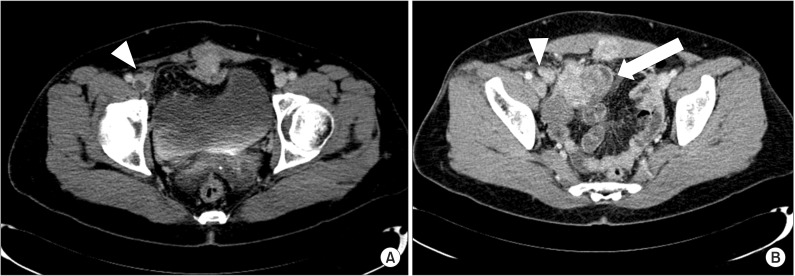
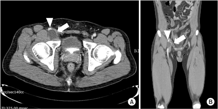
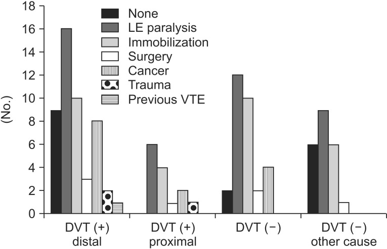
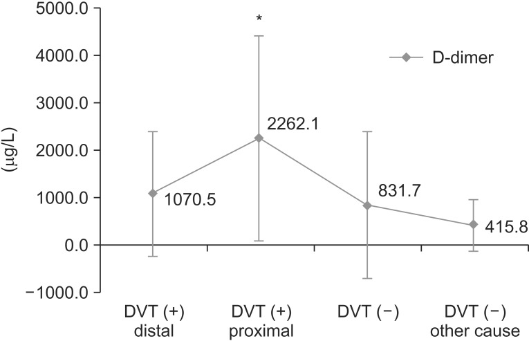
Methods: A hundred twenty-three patients, who had performed CTV performed because of suspected DVT in our clinic, were enrolled. We performed chart reviews retrospectively and categorized CTV findings as follows: DVT distal to inguinal ligament and no compression lesion; DVT proximal to inguinal ligament and no compression lesion; DVT distal to inguinal ligament and anatomical variant (for example, May-Thurner syndrome); DVT due to compression of mass (cancer or cyst); DVT and other incidental abnormal finding; and no DVT and other possible causes of leg swelling.
Results: DVTs were found in 65 (53%) patients. DVTs were found at distal level (thigh or lower leg) to inguinal ligament in 47 patients. DVTs were found at proximal to inguinal ligament, usually undetectable with duplex ultrasonography, in 6 patients. DVTs caused by external compression, such as femoral vein and cancer mass, were found in 12 patients (10%), which are also not easily detected with duplex ultrasonography. Other various causes of leg edema without DVT were found in 22 (18%) patients.
Conclusion: CTV can evaluate more extensively venous problems in the pelvis and abdomen and detect other possible causes of leg swelling. Therefore, CTV can be a useful tool not only for easy detection of DVT but also for evaluating differential diagnosis of leg edema in rehabilitation patients.
Keywords: Computed tomography; Edema; Venography; Venous thrombosis.
Conflict of interest statement
No potential conflict of interest relevant to this article was reported.
Figures
References
-
- Ely JW, Osheroff JA, Chambliss ML, Ebell MH. Approach to leg edema of unclear etiology. J Am Board Fam Med. 2006;19:148–160. - PubMed
-
- Kelly J, Rudd A, Lewis R, Hunt BJ. Venous thromboembolism after acute stroke. Stroke. 2001;32:262–267. - PubMed
-
- Byrne JJ. Phlebitis; a study of 748 cases at the Boston City Hospital. N Engl J Med. 1955;253:579–586. - PubMed
-
- Collins R, Scrimgeour A, Yusuf S, Peto R. Reduction in fatal pulmonary embolism and venous thrombosis by perioperative administration of subcutaneous heparin: overview of results of randomized trials in general, orthopedic, and urologic surgery. N Engl J Med. 1988;318:1162–1173. - PubMed
LinkOut - more resources
Full Text Sources
Other Literature Sources