Review
. 2014 Dec 15:2:72.
doi: 10.3389/fcell.2014.00072. eCollection 2014.

Mitochondrial metabolism and the control of vascular smooth muscle cell proliferation

Affiliations
Review

Mitochondrial metabolism and the control of vascular smooth muscle cell proliferation

Mario Chiong et al. Front Cell Dev Biol. .

Abstract

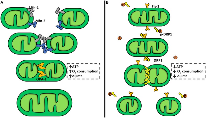
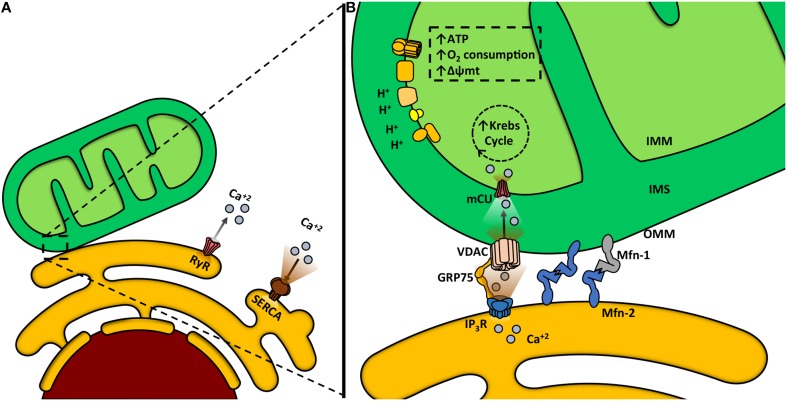
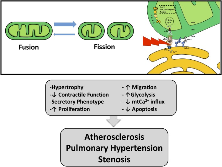
Differentiation and dedifferentiation of vascular smooth muscle cells (VSMCs) are essential processes of vascular development. VSMC have biosynthetic, proliferative, and contractile roles in the vessel wall. Alterations in the differentiated state of the VSMC play a critical role in the pathogenesis of a variety of cardiovascular diseases, including atherosclerosis, hypertension, and vascular stenosis. This review provides an overview of the current state of knowledge of molecular mechanisms involved in the control of VSMC proliferation, with particular focus on mitochondrial metabolism. Mitochondrial activity can be controlled by regulating mitochondrial dynamics, i.e., mitochondrial fusion and fission, and by regulating mitochondrial calcium handling through the interaction with the endoplasmic reticulum (ER). Alterations in both VSMC proliferation and mitochondrial function can be triggered by dysregulation of mitofusin-2, a small GTPase associated with mitochondrial fusion and mitochondrial-ER interaction. Several lines of evidence highlight the relevance of mitochondrial metabolism in the control of VSMC proliferation, indicating a new area to be explored in the treatment of vascular diseases.

Keywords: mitochondrial dynamics; mitochondrial metabolism; mitofusin-2; proliferation; vascular smooth muscle cell.


Figures

Figure 1
Figure 1
Mitochondrial dynamics. (A) Mitochondrial fusion. This is a two-step process which involves three different proteins: mitofusin-1 and 2 (Mfn-1 and Mfn-2) and optic atrophy protein-1 (OPA-1). Mfn-1 and Mfn-2 are transmembrane GTPases embedded in the outer mitochondrial membrane (OMM). The C-terminal coiled-coil region of Mfn-1 and Mfn-2 mediates tethering between mitochondria through homo- or heterotypic complexes formed between adjacent mitochondria. This interaction mediates OMM fusion. OPA-1 is a dynamin-related protein localized in the inner mitochondrial membrane (IMM), facing the intermembrane space. OPA-1 participates in the attachment and fusion of IMM. Mitochondrial fusion is associated with an increase in the mitochondrial potential (Δψm), oxygen consumption, and ATP production. (B) Mitochondrial fission. In this process participates dynamin-related protein-1 (DRP-1) and fission protein-1 (FIS-1). DRP-1 is a large GTPase found soluble in the cytosol of cells from where it shuttles onto and off mitochondria. DRP-1 assembles into spirals at division sites around the OMM to drive the fission process. In yeast, the mechanism for recruitment of DRP-1 to the mitochondria requires FIS-1, a tetratricopeptide domain protein anchored into and evenly coating the entire OMM. DRP-1 activity is inhibited by a protein kinase A (PKA) phosphorylation. Mitochondrial fission is associated with a Δψm, oxygen consumption, and ATP production decrease.
Figure 2
Figure 2
Mitochondria-endoplasmic reticulum (ER) coupling. (A) Ca2+ release from the ER to the cytosol is mediated by two channels: ryanodine receptor channel (RyR) and inositol trisphosphate receptor channel (InsP3R). The sarcoendoplasmic reticulum Ca2+ transport ATPase (SERCA) is a pump that transports Ca2+ from the cytoplasm into the ER. (B) Ca2+ transfer from the ER to the mitochondria occurs within the mitochondria-associated membranes (MAMs). Ca2+ is released from the ER to the mitochondria through the InsP3R. Glucose-regulated protein 75 (GRP75) is a mitochondrial chaperone that mediates the molecular interaction between InsP3R with the voltage-dependent anion channel (VDAC). Ca2+ import across the OMM occurs through VDAC. Ca2+ crosses the IMM through the mitochondrial Ca2+ uniporter channel (mCU) thanks to the considerable driving force represented by the negative transmembrane potential and the high Ca2+ concentration within the intermembrane space. Increased mitochondrial Ca2+ concentration activates mitochondrial dehydrogenases and enhances the oxidative phosphorylation, increasing oxygen consumption, Δψm and ATP production.
Figure 3
Figure 3
Effects of mitochondria and ER–mitochondrial coupling dysfunction. Unbalance in mitochondrial dynamics and ER–mitochondria uncoupling can lead to VSMC phenotypic switching promoting vascular pathologies such as atherosclerosis, stenosis and pulmonary hypertension.

References

    1. Ahn S. Y., Choi Y. S., Koo H. J., Jeong J. H., Park W. H., Kim M., et al. (2010). Mitochondrial dysfunction enhances the migration of vascular smooth muscles cells via suppression of Akt phosphorylation. Biochim. Biophys. Acta 1800, 275–281. 10.1016/j.bbagen.2009.09.005 - DOI - PubMed
    1. Allen A. M., Taylor J. M., Graham A. (2013). Mitochondrial (dys)function and regulation of macrophage cholesterol efflux. Clin. Sci. Lond. 124, 509–515. 10.1042/CS20120358 - DOI - PubMed
    1. Archer S. L., Gomberg-Maitland M., Maitland M. L., Rich S., Garcia J. G., Weir E. K. (2008). Mitochondrial metabolism, redox signaling, and fusion: a mitochondria-ROS-HIF-1alpha-Kv1.5 O2-sensing pathway at the intersection of pulmonary hypertension and cancer. Am. J. Physiol. Heart. Circ. Physiol. 294, H570–H578. 10.1152/ajpheart.01324.2007 - DOI - PubMed
    1. Bach D., Naon D., Pich S., Soriano F. X., Vega N., Rieusset J., et al. (2005). Expression of Mfn2, the Charcot–Marie–Tooth neuropathy type 2A gene, in human skeletal muscle: effects of type 2 diabetes, obesity, weight loss, and the regulatory role of tumor necrosis factor alpha and interleukin-6. Diabetes 54, 2685–2693. 10.2337/diabetes.54.9.2685 - DOI - PubMed
    1. Ballinger S. W., Patterson C., Knight-Lozano C. A., Burow D. L., Conklin C. A., Hu Z., et al. (2002). Mitochondrial integrity and function in atherogenesis. Circulation 106, 544–549. 10.1161/01.CIR.0000023921.93743.89 - DOI - PubMed

LinkOut - more resources