

Histotripsy of the Prostate in a Canine Model: Characterization of Post-Therapy Inflammation and Fibrosis
- PMID: 25566880
- PMCID: PMC4507352
- DOI: 10.1089/end.2014.0585
Histotripsy of the Prostate in a Canine Model: Characterization of Post-Therapy Inflammation and Fibrosis
Abstract
Introduction: Histotripsy is a nonthermal, noninvasive, pulsed ultrasound technology that homogenizes tissue within the targeted volume. From previous experiments, it appeared that the resultant fibrotic response from histotripsy was limited compared with the typical tissue response seen after thermoablation. The objective of this study was to characterize the inflammatory response and quantify patterns of collagen deposition 6 weeks after in vivo canine prostate histotripsy.
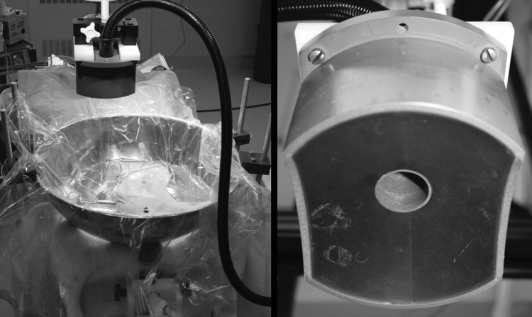
Methods: Histotripsy was applied to the left half of eight canine prostates to produce an intraparenchymal zone of tissue homogenization. Six weeks after treatment, prostates were harvested, sectioned, and stained with hematoxylin and eosin for histologic evaluation, CD3, CD20, and Mac387 immunohistochemistry to characterize the inflammatory components, and picrosirius red staining to identify collagen.
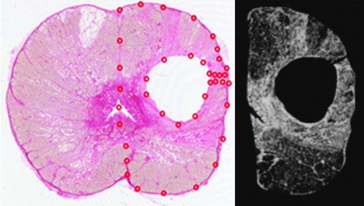
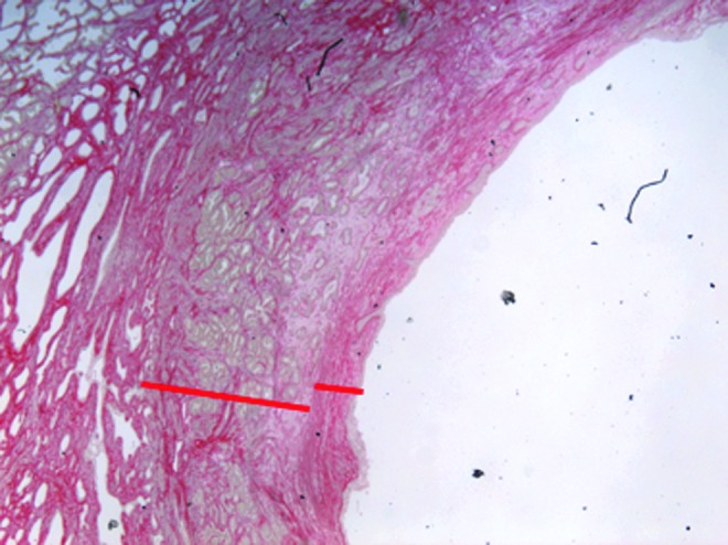
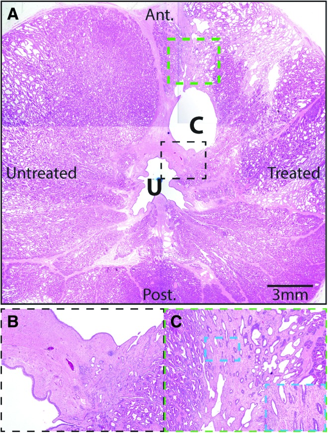
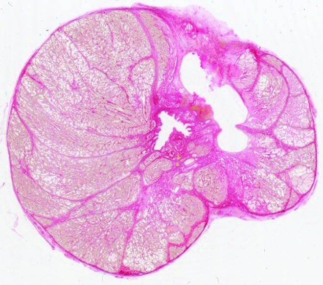
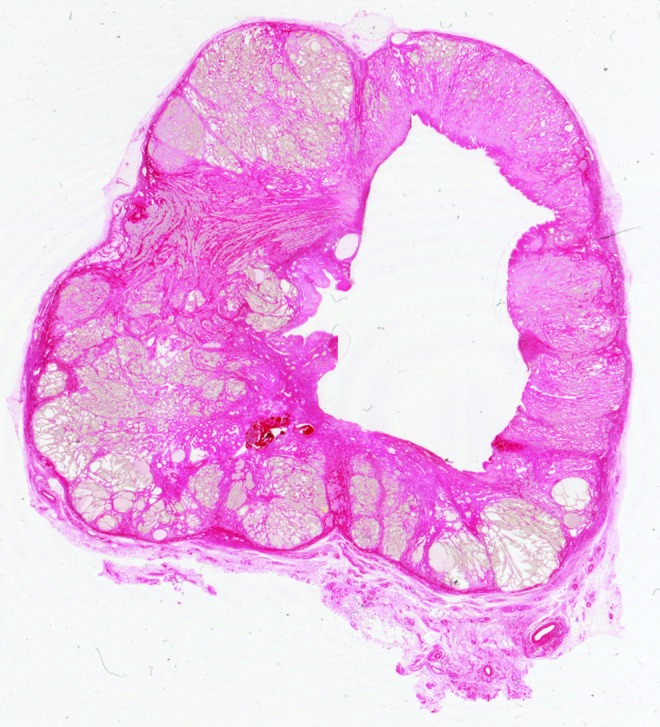
Results: Seven of eight treated prostates exhibited only minimal residual inflammation. Visual microscopic analysis of picrosirius red slides revealed a band of dense collagen (0.5 mm wide) immediately adjacent to the cavity produced by histotripsy. This was surrounded by a second band (1 mm wide) of less dense collagen interspersed among glandular architecture. A lobar distribution of epithelial atrophy and basal cell hyperplasia reminiscent of periurethral glands and ducts was apparent surrounding the margin of the treatment cavities. Tissue loss (-31%) was apparent on the treated side of all prostates while four demonstrated a net decrease in collagen content.
Conclusions: In vivo histotripsy of canine prostate produced a decrease in prostate volume coupled with a limited inflammatory and fibrotic response. A narrow (1.5 mm) band of fibrosis around the empty, reepithelialized treatment cavity was observed 6 weeks after treatment. In four cases, an overall reduction in collagen content was measured. Further studies are planned to correlate these histologic findings with alteration in mechanical tissue properties and to explore histotripsy strategies for treatment of benign prostatic hyperplasia that optimize tissue volume removal with minimization of fibrosis.
Figures
References
-
- Cantiello F, Cicione A, Salonia A, et al. Periurethral fibrosis secondary to prostatic inflammation causing lower urinary tract symptoms: A prospective cohort study. Urology 2013;81:1018–1023 - PubMed
-
- Apte M, Pirola R, Wilson J. The fibrosis of chronic pancreatitis: New insights into the role of pancreatic stellate cells. Antioxid Redox Signal 2011;15:2711–2722 - PubMed
-
- Novo E, di Bonzo LV, Cannito S, et al. Hepatic myofibroblasts: A heterogeneous population of multifunctional cells in liver fibrogenesis. Int J Biochem Cell Biol 2009;41:2089–2093 - PubMed
Publication types
MeSH terms
Substances
Grants and funding
LinkOut - more resources
Full Text Sources
Other Literature Sources
