. 2015 Jan 7;6(1):5.
doi: 10.1186/scrt539.

Bone morphogenetic protein 2 gene transduction enhances the osteogenic potential of human urine-derived stem cells

Affiliations

Bone morphogenetic protein 2 gene transduction enhances the osteogenic potential of human urine-derived stem cells

Junjie Guan et al. Stem Cell Res Ther. .

Abstract

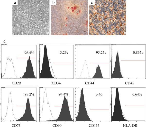
Introduction: Urine-derived stem cells (USCs) have the ability to differentiate into osteogenic lineage. Previous studies have raised the possibility that USCs could be used for bone repair. To harness the power of USCs in promoting bone regeneration, methods must be developed to induce USCs to osteogenic lineage efficiently. The present study investigates the effect of lentivirus-encoded bone morphogenetic protein 2 (BMP2) gene transduction on the osteogenic potential of USCs.

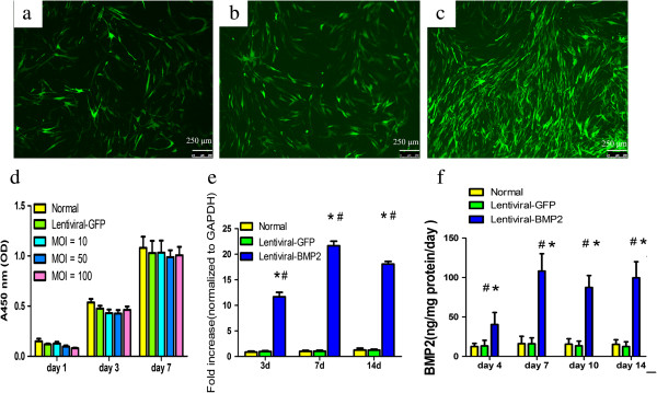
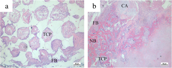
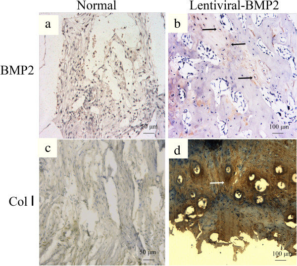
Methods: USCs were isolated from voided urine and transduced with Lentiviral vector encoding BMP2. An in vitro study was performed to detect Lentiviral-BMP2 transduced USCs differentiated towards osteogenic lineage. Furthermore, Lentiviral-BMP2 transduced USCs were transplanted in vivo to examine the ectopic bone formation ability. After six weeks, retrieval samples were obtained for immunostaining and histological analysis.

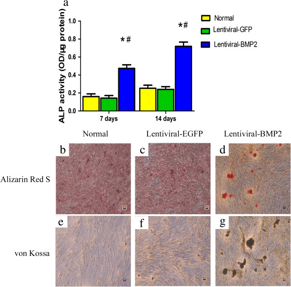
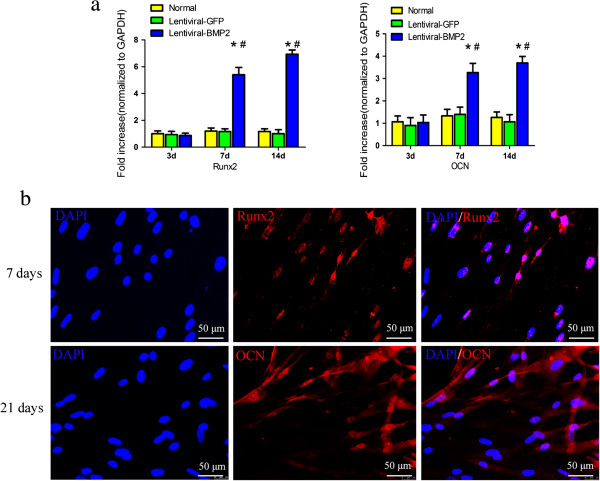
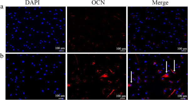
Results: The results showed that the transduction efficiencies were over 90%, and transduced USCs had high expression levels of the BMP2 gene and secreted BMP2 protein. Alkaline activity and mineral deposition staining demonstrated that transduced USCs differentiate into osteogenic lineages without the addition of osteogenic supplements. Transduced USCs also showed high expression of bone-related markers, including runt-related protein-2 (Runx2) and osteocalcin (OCN), confirming this lentiviral-BMP2 construct provides sufficient stimuli for osteogenic differentiation. Histological analysis indicated that the transduced USCs induced robust new bone formation in nude mice. Six weeks after transplantation, human derived cells were observed to participate in bone formation.

Conclusions: These results demonstrate that BMP2 gene transduction provides an effective method to enhance the osteogenic potential of USCs.


Figures

Figure 1
Figure 1
Characterization of human urine-derived stem cells. (a) Phase-contrast microscopy of USCs in primary culture. (b) Alizarin Red S staining showing the osteogenic differentiation of USCs. (c) Oil red O staining showing the adipogenic differentiation of USCs. (d). Flow cytometry analysis of the expression of USCs surface markers. Scale bars = 100 μm. USCs, urine-derived stem cells.
Figure 2
Figure 2
Lentiviral-BMP2 transduced USCs and the effect of transduction on USCs proliferation and BMP2 gene and protein expression. USCs with Lentiviral-BMP2 vectors at different MOIs (a:10; b:50; c:100). Cells were assessed for the presence of positive GFP fluorescence microscopy three days after transduction. Scale bar = 250 μm. (d) Cell proliferation assays were performed at 1, 3 and 7 days post-transduction. The means and standard deviations were calculated from three experiments. (e) RT-PCR analysis showed that the transduced USCs highly expressed the BMP2 gene at days 3, 7 and 14 after transduction. (f) ELISA results indicated that BMP2 production in Lentiviral-BMP2 transduced USCs was significantly increased as compared to that in Lentiviral-GFP transduced USCs or normal USCs. #, P <0.01 (compared with normal USCs) and *, P <0.01 (compared with Lentiviral-GFP transduced USCs). BMP2, bone morphogenic proteins 2; MOI, multiplicity of infection; USCs, urine-derived stem cells.
Figure 3
Figure 3
In vitro osteogenic differentiation of lentivirus-transduced USCs. (a) ALP activity in Lentiviral-BMP2 transduced USCs was significantly higher than that in Lentiviral-GFP transduced USCs and normal USCs. The deposited calcium was detected using Alizarin Red S (b, d and d) and von Kossa staining (e, f and g) at 14 days after transduction. (b and e) Normal USCs. (c and f) Lentiviral-GFP transduced USCs. (d and g) Lentiviral-BMP2 transduced USCs. For Alizarin Red S staining, the red color indicates calcium deposition (d); for von Kossa staining, dark patches indicate calcium deposition (g). Scale bar = 100 μm. #, P <0.05 (compared with normal USCs) and *, P <0.05 (compared with Lentiviral-GFP transduced USCs). ALP, alkaline phosphatase; BMP2, bone morphogenic proteins 2; USCs, urine-derived stem cells.
Figure 4
Figure 4
Gene and protein expression of bone related factors in transduced and normal USCs. (a) The mRNA expression level of Runx2 and OCN at day 3, 7 and 14 after transduction. (b) The protein expression of Runx2 and OCN was detected with immunofluorescence. Blue: DAPI; red: Runx2 or OCN. BMP2 gene transduction significantly increased the protein expression of Runx2 and OCN. Scale bar = 50 μm. #, P <0.05 (compared with normal USCs) and *, P <0.05 (compared with Lentiviral-GFP transduced USCs). BMP2, bone morphogenic proteins 2; DAPI, 4',6-diamidino-2-phenylindole; OCN, osteocalcin; Runx2, Runt-related protein-2; USCs, urine-derived stem cells.
Figure 5
Figure 5
H & E staining of histological sections formed by the implantation of Lentiviral-BMP2 transduced USCs/β-TCP and USCs/β-TCP into the hind limb of athymic mice for six weeks. (a) The USCs/β-TCP induced no bone formation, whereas the Lentiviral-BMP2 transduced USCs/β-TCP resulted in bone formation in the outer surface and inner pore of the implants (b). Scale bar = 50 μm. TCP: β-TCP; FB: fibroblast tissue; NB: new bone; CA: cartilage tissue. B-TCP, β-tricalcium phosphate; BMP2, bone morphogenic proteins 2; USCs, urine-derived stem cells.
Figure 6
Figure 6
Immunohistochemical staining of BMP2 and Collagen I of the transplanted scaffold at six weeks post-implantation. (a) BMP2 and (c) Collagen I staining in the normal USCs transplantation group. (b) BMP2 and (d) Collagen I staining in the Lentiviral-BMP2 transplantation group. The arrows indicate the positive staining of BMP2 or Collagen I in the Lentiviral-BMP2 transplantation group. Scale bar for a and c = 50 μm; b and d = 100 μm. BMP2, bone morphogenic proteins 2; USCs, urine-derived stem cells.
Figure 7
Figure 7
Human derived OCN was detected by immunostaining after six weeks. (a) USCs/β-TCP; (b) Lentiviral-BMP2 transduced USCs/β-TCP. The arrows indicate the positive staining of OCN. Scale bar = 100 μm. B-TCP, β-tricalcium phosphate; BMP2, bone morphogenic proteins 2; OCN, osteocalcin; USCs, urine-derived stem cells.

References

    1. Zhang Y, McNeill E, Tian H, Soker S, Andersson KE, Yoo JJ, et al. Urine derived cells are a potential source for urological tissue reconstruction. J Urol. 2008;180:2226–33. doi: 10.1016/j.juro.2008.07.023. - DOI - PubMed
    1. Guan J, Niu X, Gong F, Hu B, Guo S, Lou Y, et al. Biological characteristics of human urine derived stem cells: potential for cell-based therapy in neurology. Tissue Eng Part A. 2014;6:6. - PMC - PubMed
    1. Bodin A, Bharadwaj S, Wu S, Gatenholm P, Atala A, Zhang Y. Tissue-engineered conduit using urine-derived stem cells seeded bacterial cellulose polymer in urinary reconstruction and diversion. Biomaterials. 2010;31:8889–901. doi: 10.1016/j.biomaterials.2010.07.108. - DOI - PubMed
    1. Wu S, Liu Y, Bharadwaj S, Atala A, Zhang Y. Human urine-derived stem cells seeded in a modified 3D porous small intestinal submucosa scaffold for urethral tissue engineering. Biomaterials. 2011;32:1317–26. doi: 10.1016/j.biomaterials.2010.10.006. - DOI - PubMed
    1. Bishop GB, Einhorn TA. Current and future clinical applications of bone morphogenetic proteins in orthopaedic trauma surgery. Int Orthop. 2007;31:721–7. doi: 10.1007/s00264-007-0424-8. - DOI - PMC - PubMed

Publication types

MeSH terms