. 2009 Feb;2(1):101-12.
doi: 10.1111/j.1752-4571.2008.00064.x.

Heart disease is common in humans and chimpanzees, but is caused by different pathological processes

Affiliations

Heart disease is common in humans and chimpanzees, but is caused by different pathological processes

Nissi Varki et al. Evol Appl. 2009 Feb.

Abstract

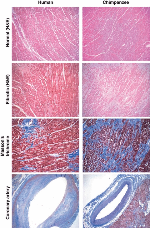
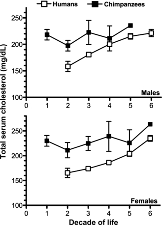
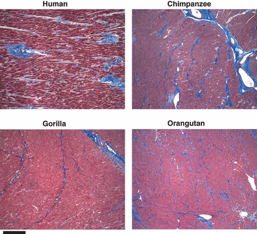
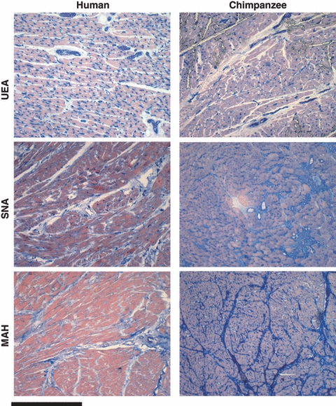
Heart disease is common in both humans and chimpanzees, manifesting typically as sudden cardiac arrest or progressive heart failure. Surprisingly, although chimpanzees are our closest evolutionary relatives, the major cause of heart disease is different in the two species. Histopathology data of affected chimpanzee hearts from two primate centers, and analysis of literature indicate that sudden death in chimpanzees (and in gorillas and orangutans) is commonly associated with diffuse interstitial myocardial fibrosis of unknown cause. In contrast, most human heart disease results from coronary artery atherosclerosis, which occludes myocardial blood supply, causing ischemic damage. The typical myocardial infarction of humans due to coronary artery thrombosis is rare in these apes, despite their human-like coronary-risk-prone blood lipid profiles. Instead, chimpanzee 'heart attacks' are likely due to arrythmias triggered by myocardial fibrosis. Why do humans not often suffer from the fibrotic heart disease so common in our closest evolutionary cousins? Conversely, why do chimpanzees not have the kind of heart disease so common in humans? The answers could be of value to medical care, as well as to understanding human evolution. A preliminary attempt is made to explore possibilities at the histological level, with a focus on glycosylation changes.

Keywords: atherosclerosis; evolution; great ape; heart attacks; heart disease; heart failure; hominids; myocardial fibrosis.


Figures

Figure 1
Figure 1
Examples of histological comparisons of human and chimpanzee hearts and coronary blood vessels. Top panels: Hematoxylin and eosin stains of normal myocardium sections from humans and chimpanzees look very similar. Upper middle panels: Example of fibrosis immediately surrounding blood vessels, as seen in some human hearts, and example of extensive interstitial myocardial fibrosis in chimpanzee heart. Lower middle panels: Collagen fibrosis is more clearly seen in Masson's trichrome stain. Bottom panels: Atherosclerotic coronary artery typically seen in humans (note the subendothelial plaques), compared with a typical uninvolved coronary artery in a chimpanzee, as seen in Masson's Trichrome stain.
Figure 2
Figure 2
Longitudinal age-based comparison of total serum cholesterol in human and captive chimpanzee populations. Chimpanzee and human cholesterol levels were taken from two independent, original articles and plotted here, on the same axis, for comparison. Total serum cholesterol data from both publications was grouped by sex and age in decades. Human data were acquired from 6757 participants in the Framingham Heart Study, aged 15–79 years (Abbott et al. 1983). Chimpanzee data were acquired from 252 chimpanzees, aged 0–60, living in captivity at the Yerkes Primate Center (Herndon and Tigges 2001). Data are plotted as mean ± 95% CI. Error bars are not shown for the oldest chimps (5th decade in males, n = 1; 6th decade in females, n = 3) due to the very small sample size. Note that the total serum cholesterol (Y axis) starts at 100 mg/dL, to make the differences clearer.
Figure 3
Figure 3
Masson's trichrome staining for collagen in histologically normal-appearing myocardial tissue sections from humans and great apes. ×40 magnification. Scale bar represents 50 microns. Examples are shown of the staining patterns in human and great apes. The results shown are typical of that seen with six human, six chimpanzee, three gorilla, and four orangutan samples.
Figure 4
Figure 4
Lectin staining of histologically normal-appearing myocardial sections from humans and great apes. Examples are shown of the typical lectin staining patterns (blue) in human and chimpanzee myocardial sections with Ulex Europaeus Agglutinin (UEA), which recognizes terminal fucose residues in the sequence Fucα1-2Galβ1-4GlcNAcβ-; Sambucus nigra agglutinin (SNA) which recognizes terminal Siaα2-6Galβ1-4GlcNAcβ- units on N-linked glycan chains of glycoproteins; and, Maackia Amurensis hemagglutinin (MAH), which recognizes Siaα2-3Gal termini on various glycoconjugates (Martin et al. 2002; Varki and Varki 2007). The results shown are typical of that seen with 11 human and seven chimpanzee samples. Three gorilla and four orangutan samples gave results similar to that of the chimpanzee sections.

References

    1. Abbott RD, Garrison RJ, Wilson PW, Epstein FH, Castelli WP, Feinleib M, LaRue C. Joint distribution of lipoprotein cholesterol classes. The Framingham study. Arteriosclerosis. 1983;3:260–272. - PubMed
    1. Altenburg M, Johnson L, Wilder J, Maeda N. Apolipoprotein E4 in macrophages enhances atherogenesis in a low density lipoprotein receptor-dependent manner. Journal of Biological Chemistry. 2007;282:7817–7824. - PMC - PubMed
    1. Binder CJ, Chang MK, Shaw PX, Miller YI, Hartvigsen K, et al. Innate and acquired immunity in atherogenesis. Nature Medicine. 2002;8:1218–1226. - PubMed
    1. Binder CJ, Shaw PX, Chang MK, Boullier A, Hartvigsen K, et al. The role of natural antibodies in atherogenesis. Journal of Lipid Research. 2005;46:1353–1363. - PubMed
    1. Blaton V, Peeters H. The nonhuman primates as models for studying human atherosclerosis: studies on the chimpanzee, the baboon and the rhesus macacus. Advances in Experimental Medicine and Biology. 1976;67:33–64. - PubMed