. 2015 Jan;43(2):862-74.
doi: 10.1093/nar/gku1396. Epub 2015 Jan 7.

A composite enhancer regulates p63 gene expression in epidermal morphogenesis and in keratinocyte differentiation by multiple mechanisms

Affiliations

A composite enhancer regulates p63 gene expression in epidermal morphogenesis and in keratinocyte differentiation by multiple mechanisms

Dario Antonini et al. Nucleic Acids Res. 2015 Jan.

Abstract

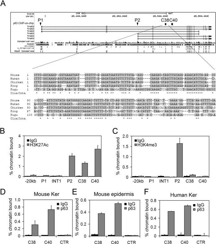
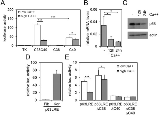
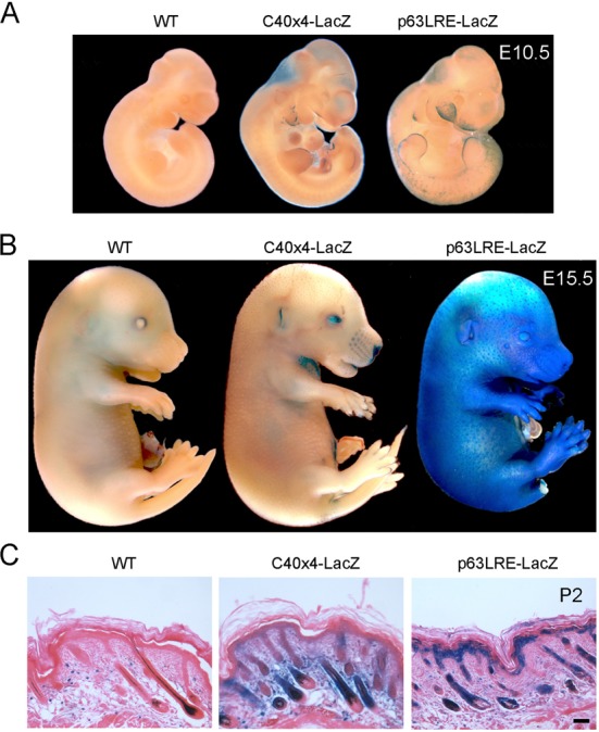
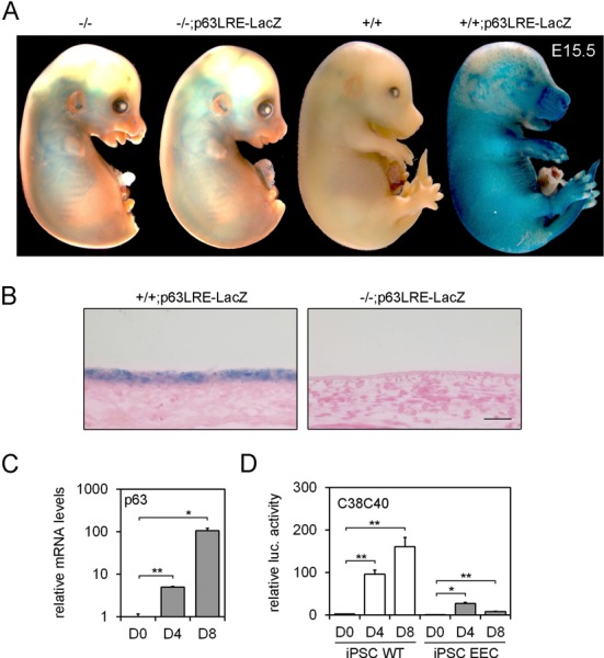
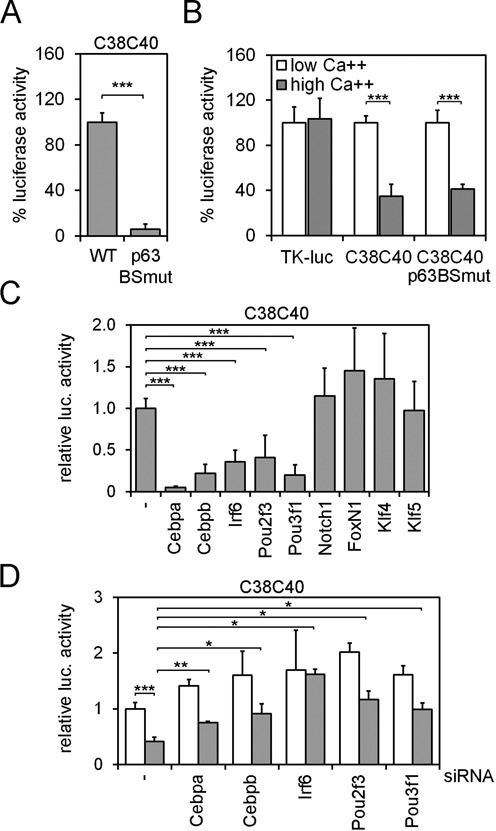
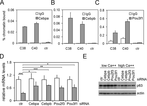
p63 is a crucial regulator of epidermal development, but its transcriptional control has remained elusive. Here, we report the identification of a long-range enhancer (p63LRE) that is composed of two evolutionary conserved modules (C38 and C40), acting in concert to control tissue- and layer-specific expression of the p63 gene. Both modules are in an open and active chromatin state in human and mouse keratinocytes and in embryonic epidermis, and are strongly bound by p63. p63LRE activity is dependent on p63 expression in embryonic skin, and also in the commitment of human induced pluripotent stem cells toward an epithelial cell fate. A search for other transcription factors involved in p63LRE regulation revealed that the CAAT enhancer binding proteins Cebpa and Cebpb and the POU domain-containing protein Pou3f1 repress p63 expression during keratinocyte differentiation by binding the p63LRE enhancer. Collectively, our data indicate that p63LRE is composed of additive and partly redundant enhancer modules that act to direct robust p63 expression selectively in the basal layer of the epidermis.


Figures

Figure 1.
Figure 1.
(A) Upper panel: mouse p63 gene structure. The P1 (TA) and P2 (ΔN) promoters are indicated. C38 and C40 correspond to highly conserved sequences in evolution as determined by the 17-way alignment predicted by the phastCons program (56). Genomic elements identified by ChIP-on-chip experiment using p63 antibodies are indicated with black squares (25). Lower panel: evolutionary conservation of the C38 element in multiple vertebrate species. Conserved nucleotides are shown in gray, and identical nucleotides in all species are indicated by stars (Clustal consensus). (B,C) ChIP-qPCR was performed on mouse primary keratinocyte using antibodies anti-H3K27Ac (B), anti-H3K4me3 (C) (gray bars) or rabbit IgG (black bars) as negative control. (D,E,F) ChIP-qPCR was performed on primary mouse keratinocytes (D), mouse newborn epidemis (E) and human keratinocytes (F) using p63 polyclonal antibodies (H-137) (gray bars), or rabbit IgG (black bars) as negative control. Oligonucleotide sequences are listed in Supplementary Data. Error bars denote SD.
Figure 2.
Figure 2.
(A) C38-Luc, C40-Luc and C38C40-Luc enhancer activity was analyzed in primary mouse keratinocytes under proliferation (low calcium; white bars) and differentiation conditions (high calcium; gray bars) upon 24 h of calcium treatment (Ca++ 2 mM). Data are expressed relative to TK promoter activity used as control (*P ≤ 0.05; ***P ≤ 0.0005; n = 4). (B,C) p63 expression was detected at the mRNA (B) and protein (C) levels in primary mouse keratinocytes in proliferating and in calcium-induced differentiated keratinocytes at the indicated times (Ca++ 2 mM). (D) p63LRE luciferase activity was tested in mouse dermal fibroblasts (Fib) and primary keratinocytes (Ker). (E) TK-Luc construct carrying p63LRE, p63LREΔC38, p63LREΔC40 and p63LREΔC38ΔC40 were tested for luciferase activity in primary mouse keratinocytes under proliferation (white bars) and differentiation (gray bars) conditions (***P ≤ 0.0005; n = 6). Error bars denote SD.
Figure 3.
Figure 3.
(A) E10.5 wild type (WT) and transgenic embryos containing four copies of C40 (C40×4-LacZ) or p63LRE-LacZ were tested for β-galactosidase activity. (B) Embryos at E15.5 were tested for β-galactosidase activity as in (A). (C) Histological sections of skin at P2 were processed for β-galactosidase staining and counterstained with eosin. Scale bar, 50 μm.
Figure 4.
Figure 4.
(A) E15.5 embryos carrying p63LRE in a wild type (+/+; p63LRE-LacZ) and in a p63-null background (-/-; p63LRE-LacZ) were tested for β-galactosidase activity. Wild-type (+/+) and p63-null (-/-) littermates were used as controls. (B) Histological sections of skin at E15.5 were obtained after β-galactosidase staining and were subsequently paraffin embedded and counterstained with eosin. Scale bar, 50 μm. (C) p63 expression was detected at the mRNA levels in human iPSCs at 0, 4 and 8 days (D0–D8) of corneal epithelial differentiation. Data are represented relative to p63 expression levels at day 0 (*P ≤ 0.05; **P ≤ 0.005; n = 3). (D) Luciferase activity of C38C40-Luc was tested in human iPSCs (WT and EEC) at D0–D8 of corneal epithelial differentiation. Data are represented relative to luciferase activity of TK control in D0 human iPSCs (*P ≤ 0.05; **P ≤ 0.005; n = 3).
Figure 5.
Figure 5.
(A) Luciferase activity of the C38C40-Luc carrying either WT or mutant p63-binding sites (p63BSmut) was tested in mouse keratinocytes. Data are expressed as the% of wild type luciferase activity (***P ≤ 0.0005; n = 3). (B) The activity of wild type C38C40, C38C40p63BSmut and TK control was tested in proliferating (low Ca++; white bars) and differentiated keratinocytes (2 mM Ca++; gray bars), and plotted as% of each luciferase activity in low Ca++ conditions. (***P ≤ 0.0005; n = 3). (C) C38C40-Luc luciferase activity in the absence (−) or in the presence of expression vectors carrying the indicated TFs in mouse keratinocytes. (***P ≤ 0.0005; n = 7). (D) C38C40 luciferase activity was tested in differentiated (gray bars) and proliferating (white bars) mouse keratinocytes upon transfection of the indicated siRNAs. (*P ≤ 0.05; **P ≤ 0.005; ***P ≤ 0.0005; n = 3). Error bars denote SD.
Figure 6.
Figure 6.
(A, B, C) ChIP-qPCR was performed on mouse primary keratinocytes using antibodies specific for Cebpa (A), Cebpb (B), Pou3f1 (C) (gray bars) or rabbit IgG as negative control (white bars). (D) p63 mRNA expression levels in differentiated (gray bars) and proliferating (white bars) keratinocytes upon transfection of the indicated siRNAs. (*P ≤ 0.05; **P ≤ 0.005; ***P ≤ 0.0005; n = 4). Error bars denote SD. (E) Immunoblotting of total cell extracts from primary mouse keratinocytes transfected as in (D) using antibodies against the indicated proteins. Keratinocytes were cultured in proliferation (low Ca++) or in differentiation conditions (0.6 mM Ca++ for 24 h).

References

    1. Mills A.A., Zheng B., Wang X.J., Vogel H., Roop D.R., Bradley A. p63 is a p53 homologue required for limb and epidermal morphogenesis. Nature. 1999;398:708–713. - PubMed
    1. Yang A., Schweitzer R., Sun D., Kaghad M., Walker N., Bronson R.T., Tabin C., Sharpe A., Caput D., Crum C., et al. p63 is essential for regenerative proliferation in limb, craniofacial and epithelial development. Nature. 1999;398:714–718. - PubMed
    1. Ferone G., Thomason H.A., Antonini D., De Rosa L., Hu B., Gemei M., Zhou H., Ambrosio R., Rice D.P., Acampora D., et al. Mutant p63 causes defective expansion of ectodermal progenitor cells and impaired FGF signalling in AEC syndrome. EMBO Mol. Med. 2012;4:192–205. - PMC - PubMed
    1. Pellegrini G., Dellambra E., Golisano O., Martinelli E., Fantozzi I., Bondanza S., Ponzin D., McKeon F., De Luca M. p63 identifies keratinocyte stem cells. Proc. Natl. Acad. Sci. U.S.A. 2001;98:3156–3161. - PMC - PubMed
    1. Senoo M., Pinto F., Crum C.P., McKeon F. p63 Is essential for the proliferative potential of stem cells in stratified epithelia. Cell. 2007;129:523–536. - PubMed

Publication types