Review
. 2015 Jan;3(1):1-11.
doi: 10.1158/2326-6066.CIR-14-0209.

Tumors: wounds that do not heal-redux

Affiliations
Review

Tumors: wounds that do not heal-redux

Harold F Dvorak. Cancer Immunol Res. 2015 Jan.

Abstract

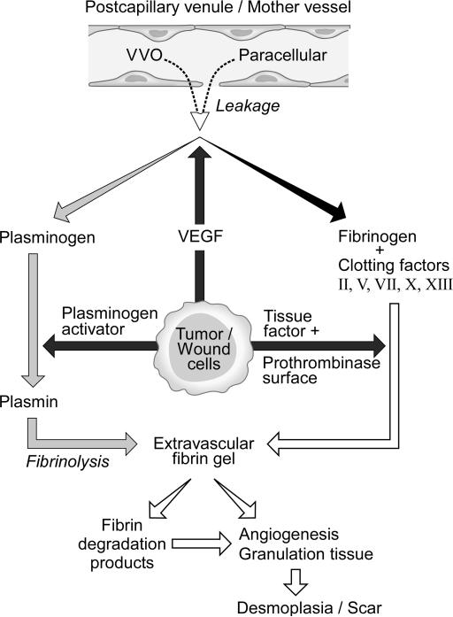
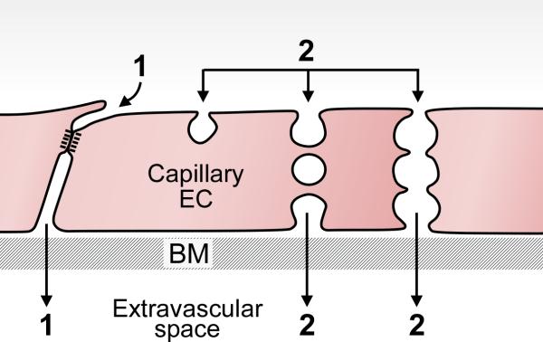
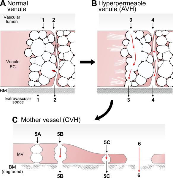
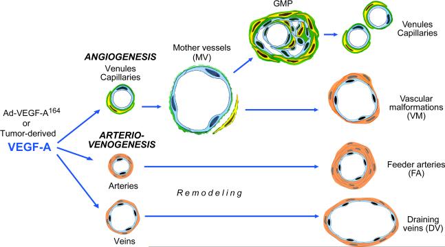
Similarities between tumors and the inflammatory response associated with wound healing have been recognized for more than 150 years and continue to intrigue. Some years ago, based on our then recent discovery of vascular permeability factor (VPF)/VEGF, I suggested that tumors behaved as wounds that do not heal. More particularly, I proposed that tumors co-opted the wound-healing response to induce the stroma they required for maintenance and growth. Work over the past few decades has supported this hypothesis and has put it on a firmer molecular basis. In outline, VPF/VEGF initiates a sequence of events in both tumors and wounds that includes the following: increased vascular permeability; extravasation of plasma, fibrinogen and other plasma proteins; activation of the clotting system outside the vascular system; deposition of an extravascular fibrin gel that serves as a provisional stroma and a favorable matrix for cell migration; induction of angiogenesis and arterio-venogenesis; subsequent degradation of fibrin and its replacement by "granulation tissue" (highly vascular connective tissue); and, finally, vascular resorption and collagen synthesis, resulting in the formation of dense fibrous connective tissue (called "scar tissue" in wounds and "desmoplasia" in cancer). A similar sequence of events also takes place in a variety of important inflammatory diseases that involve cellular immunity.


Figures

Figure 1
Figure 1
Schematic diagram of stroma formation in tumors and wounds. Stroma formation is initiated by tumor or wound cell expression of VEGF that leads to vascular hyperpermeability and consequent plasma protein extravasation, fibrin deposition and generation of highly vascular immature and then mature connective tissue stroma.
Figure 2
Figure 2
Schematic diagram illustrating pathways by which molecules of varying sizes cross the normal capillary barrier. Path 1, paracellular pathway through inter-endothelial cell cleft for small molecule extravasation. This pathway is closed to large molecules such as plasma proteins by specialized adherens and occludens junctions. Path 2, caveolae may shuttle across the capillary endothelium or form a chain of vesicles that connect the lumen and albumen to provide a pathway for plasma protein extravasation. BM, basement membrane. Modified from (43).
Figure 3
Figure 3
(A) Schematic diagram of a normal venule comprising cuboidal endothelial cells with prominent VVOs and a zone of tight apposition representing occludens and adherens junctions as in Fig. 2. Some VVO vesicles attach to the intercellular cleft, above or below (arrow) specialized junctions. Paths 1 and 2 indicate potential pathways for transcellular (VVO) and paracellular (intercellular) plasma extravasation, respectively. Basement membrane (BM) is intact and the endothelium is covered by pericytes (not shown). (B) AVH. Acute exposure to VEGF-A causes VVOs to open, allowing transcellular passage of plasma proteins (Path 3), possibly by mechanical pulling apart of stomatal diaphragms. Plasma extravasation may also take place through opened intercellular junctions (Path 4). (C) CVH. Prolonged VEGF-A stimulation causes venules to transform into mother vessels. Plasma may extravasate either through residual VVO vesicles (Path 5) or through fenestrae (Path 6). Modified from (43).
Figure 4
Figure 4
A. Electron micrograph illustrating a cross section of a normal venule with typical prominent VVOs in the cytoplasm. B. An ultrathin section of a single VVO, typically located adjacent to the intercellular cleft that separates adjacent endothelial cells. Three sequences of interconnecting vesicles-vacuoles (a,b,c) form transendothelial chains as observed in this and subsequent serial sections. C. Computer-generated three-dimensional reconstruction of portion of a VVO, extending from the lumen (bottom, with two openings indicated by arrow) to the ablumen (top, four openings were identified but are not shown in this projection). D. Electron micrograph of a venule in skin following local injection of VEGF and iv injection of colloidal carbon. Open arrowheads indicate five separate carbon-filled, trans-endothelial cell pores that pass through adjacent endothelial cells. The intercellular cleft and occludens-type junctions (solid arrows) between these two apposed cells remain intact. E. Typical, mother vessel (MV) with thinned cytoplasm, enlarged lumen filled with red blood cells, and detached pericytes. Arrow points to a mitotic figure. Inset: the normal venule in A is reproduced at the same magnification as the MV to illustrate differences in relative size of normal venules and MV. F. VVO of a MV supplying a mouse tumor is filled with reaction product 10 seconds after iv injection of tracer horseradish peroxidase. Reaction product is confined to VVO vesicles that extend from lumen (L) to ablumen (open arrowhead). Intercellular cleft (solid arrowhead) contains no peroxidase reaction product, providing definitive evidence that protein tracer passed preferentially through the cell via VVOs rather than by a paracellular route between endothelial cells. G. MV endothelium is extremely thinned and spanned by few residual vesicles, one of which (arrowhead) traverses cytoplasm to touch both luminal and abluminal plasma membrane. Another (solid arrow) forms deep abluminal invagination. Open arrows indicate intercellular cleft which is closed and not able to accommodate circulating ferritin tracer. H. Fenestrated portion of MV endothelium in a mouse tumor following iv injection of 150kDa fluoresceinated dextran. Dextran particles are visualized in vascular lumen and immediately above and below fenestrae with diaphragms (solid arrows), and abundantly in the underlying basal lamina. One fenestra (open arrow) contains dextran particles and lacks a visible diaphragm. R, red blood cell; L, vascular lumen. Bar, 200 nm. Panels were produced from references, as follows: A, E, G, (67). B, C, (47). D, (50). F, (46). H, (49).
Figure 5
Figure 5
Schematic diagram of the angiogenic and arterio-venogenic responses induced by VEGF in mouse tissues.

References

    1. Dvorak HF. Tumors: wounds that do not heal. Similarities between tumor stroma generation and wound healing. N Engl J Med. 1986;315:1650–9. - PubMed
    1. Balkwill F, Mantovani A. Inflammation and cancer: back to Virchow? Lancet. 2001;357:539–45. - PubMed
    1. Coussens LM, Werb Z. Inflammation and cancer. Nature. 2002;420:860–7. - PMC - PubMed
    1. Mantovani A, Allavena P, Sica A, Balkwill F. Cancer-related inflammation. Nature. 2008;454:436–44. - PubMed
    1. Sun B, Karin M. Inflammation and liver tumorigenesis. Front Med. 2013;7:242–54. - PubMed

Publication types