. 2015 Feb 3;112(5):1259-64.
doi: 10.1073/pnas.1419325112. Epub 2015 Jan 7.

A chemiosmotic mechanism of symport

Affiliations

A chemiosmotic mechanism of symport

H Ronald Kaback. Proc Natl Acad Sci U S A. .

Abstract

Lactose permease (LacY), a paradigm for the largest family of membrane transport proteins, catalyzes the coupled translocation of a galactoside and an H(+) across the Escherichia coli membrane (galactoside/H(+) symport). Initial X-ray structures reveal N- and C-terminal domains, each with six largely irregular transmembrane helices surrounding an aqueous cavity open to the cytoplasm. Recently, a structure with a narrow periplasmic opening and an occluded galactoside was obtained, confirming many observations and indicating that sugar binding involves induced fit. LacY catalyzes symport by an alternating access mechanism. Experimental findings garnered over 45 y indicate the following: (i) The limiting step for lactose/H(+) symport in the absence of the H(+) electrochemical gradient (∆µ̃H+) is deprotonation, whereas in the presence of ∆µ̃H+, the limiting step is opening of apo LacY on the other side of the membrane; (ii) LacY must be protonated to bind galactoside (the pK for binding is ∼10.5); (iii) galactoside binding and dissociation, not ∆µ̃H+, are the driving forces for alternating access; (iv) galactoside binding involves induced fit, causing transition to an occluded intermediate that undergoes alternating access; (v) galactoside dissociates, releasing the energy of binding; and (vi) Arg302 comes into proximity with protonated Glu325, causing deprotonation. Accumulation of galactoside against a concentration gradient does not involve a change in Kd for sugar on either side of the membrane, but the pKa (the affinity for H(+)) decreases markedly. Thus, transport is driven chemiosmotically but, contrary to expectation, ∆µ̃H+ acts kinetically to control the rate of the process.

Keywords: MFS; X-ray crystal structure; conformational change; membrane proteins; transport.


Conflict of interest statement

The author declares no conflict of interest.

Figures

Fig. 1.
Fig. 1.
LacY ribbon presentation in an inward-open conformation with a twofold axis of symmetry (broken line). (Left) N-terminal helix bundle (light yellow). (Right) C-terminal helix bundle (tan). The cytoplasmic side is shown at the top. The blue region represents the hydrophilic cavity, and the gray-shaded area represents the membrane.
Fig. 2.
Fig. 2.
Trp replacements in two pairs of Gly-Gly residues that connect the N- and C-terminal six-helix domains on the periplasmic side of LacY. The 12 transmembrane helices that make up LacY are colored light yellow [N-terminal (N-term) bundle] and tan [C-terminal (C-term) bundle]. Gly residues G159 and G370 in helices V and XI, respectively, and Trp replacements G46W (helix II) and G262W (helix VIII) are indicated. The putative outward-open structure is viewed from the side (A) or from the periplasm (B). The crystal structure of the almost occluded, narrow outward-open conformer of LacY with Gly→Trp replacements at positions 46 and 262 and bound galactoside (dark gray) is viewed from the side (C) or the periplasm (D), respectively.
Fig. 3.
Fig. 3.
Surface renditions of LacY G46W/G262W molecule A. (A) View from the periplasmic side showing TDG (green and red spheres) just visible within the molecule. Trp residues are shown in blue. (B) Slab view. (C) View from the cytoplasmic side with the residues that form a zipper-like motif to seal that side.
Fig. 4.
Fig. 4.
Electron density map contoured at 1σ (green mesh) of the sugar-binding site of LacY G46W/G262W. The density is superimposed on the structure, which is shown as sticks, with carbon atoms in gold, oxygen atoms in red, and nitrogen atoms in blue. Broken lines represent putative H bonds.
Fig. 5.
Fig. 5.
Cytoplasmic view of the active site in double-Trp LacY. TDG is shown as green sticks, and side chains forming H bonds with TDG are shown in yellow. Broken lines represent likely H bonds. Ala122 and Cys148, which are close to TDG but do not make direct contact, are shown in cyan. Glu325 and Arg302 are shown in purple. The green felt-like area represents the Van der Waals lining of the cavity. Note the narrow opening on the periplasmic side.
Fig. 6.
Fig. 6.
Crystal structure of single-Cys122 LacY with covalently bound MTS-Gal. (A) Side chains are shown as sticks. The yellow side chains (Glu269 and Trp151) make direct contact with the galactopyranosyl ring of MTS-Gal covalently bound to a Cys at position 122. The gray side chains are not sufficiently close to make contact with the galactopyranosyl ring. Glu325 and Arg302 (in purple) are involved in H+ transport. The green felt-like area represents the Van der Waals lining of the cavity. Note that the periplasmic side is closed. (B) Structure of single-Cys122 LacY with covalently bound MTS-Gal viewed from the side. Helices are depicted as rods, and MTS-Gal is shown as spheres colored by atom type with carbon in green. The aqueous central cavity open to the cytoplasmic side is colored light green.
Fig. 7.
Fig. 7.
Effect of pH on the apparent Kd (Kdapp) for TDG binding to WT LacY (black) and the E325A mutant (green).
Fig. 8.
Fig. 8.
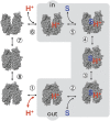
Kinetic scheme for galactoside/H+ symport, exchange, and counterflow. Symport starts with protonation of LacY (step 1 or 6 for influx or effux, respectively), which is required for high-affinity binding of lactose. Sugar (S) binding to protonated LacY (step 2 or 5) causes a conformational change to an occluded state (step 3 or 4), which can relax to either side where sugar dissociates first (step 2 or 5), followed by deprotonation (step 1 or 6) and return of unloaded LacY via an apo occluded intermediate (steps 7 and 8). Exchange or counterflow involves only steps 2–5 (gray shaded area). Because LacY catalyzes symport in both directions, when symport is in the influx direction (step 1, protonation), the pK is very alkaline (∼10.5), and step 6 (deprotonation) must have a much lower pK for deprotonation to occur (i.e., Arg302 approximates protonated Glu325). However, in the efflux direction, the pKs of these steps are reversed.

Similar articles

Cited by

References

    1. Guan L, Kaback HR. Lessons from lactose permease. Annu Rev Biophys Biomol Struct. 2006;35:67–91. - PMC - PubMed
    1. Madej MG, Kaback HR. 2014. The life and times of Lac permease: Crystals ain’t enough, but they certainly do help. Membrane Transporter Function: To Structure and Beyond, Springer Series in Biophysics: Transporters, eds Ziegler C, Kraemer R (Springer, Heidelberg) Vol 17, pp 121–158.
    1. Abramson J, et al. Structure and mechanism of the lactose permease of Escherichia coli. Science. 2003;301(5633):610–615. - PubMed
    1. Mirza O, Guan L, Verner G, Iwata S, Kaback HR. Structural evidence for induced fit and a mechanism for sugar/H+ symport in LacY. EMBO J. 2006;25(6):1177–1183. - PMC - PubMed
    1. Guan L, Mirza O, Verner G, Iwata S, Kaback HR. Structural determination of wild-type lactose permease. Proc Natl Acad Sci USA. 2007;104(39):15294–15298. - PMC - PubMed

Publication types

Substances