. 2015 Jan 7;35(1):84-95.
doi: 10.1523/JNEUROSCI.1737-14.2015.

Temporal plasticity involved in recovery from manual dexterity deficit after motor cortex lesion in macaque monkeys

Affiliations

Temporal plasticity involved in recovery from manual dexterity deficit after motor cortex lesion in macaque monkeys

Yumi Murata et al. J Neurosci. .

Abstract

The question of how intensive motor training restores motor function after brain damage or stroke remains unresolved. Here we show that the ipsilesional ventral premotor cortex (PMv) and perilesional primary motor cortex (M1) of rhesus macaque monkeys are involved in the recovery of manual dexterity after a lesion of M1. A focal lesion of the hand digit area in M1 was made by means of ibotenic acid injection. This lesion initially caused flaccid paralysis in the contralateral hand but was followed by functional recovery of hand movements, including precision grip, during the course of daily postlesion motor training. Brain imaging of regional cerebral blood flow by means of H2 (15)O-positron emission tomography revealed enhanced activity of the PMv during the early postrecovery period and increased functional connectivity within M1 during the late postrecovery period. The causal role of these areas in motor recovery was confirmed by means of pharmacological inactivation by muscimol during the different recovery periods. These findings indicate that, in both the remaining primary motor and premotor cortical areas, time-dependent plastic changes in neural activity and connectivity are involved in functional recovery from the motor deficit caused by the M1 lesion. Therefore, it is likely that the PMv, an area distant from the core of the lesion, plays an important role during the early postrecovery period, whereas the perilesional M1 contributes to functional recovery especially during the late postrecovery period.

Keywords: brain activation; functional compensation; macaque monkey; precision grip; primate.


Figures

Figure 1.
Figure 1.
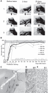
Recovery of precision grip after lesion of M1. A, Sequence of photographs showing hand and digit movements while performing the precision grip task (Monkey [Mk]-Ja), in which monkeys grasped a small cubic knob attached to a plastic disk and retrieved it from a tube through a slit aperture. The joints and tips of the thumb and index finger are linked by solid and dotted lines, respectively. Rows 1, 2, and 3 show the moment of contact of the tips of the index finger with the aperture of the slot, contact between the tip of the index finger and the square knob, and retrieval of the object, respectively. B, Time course of changes in the success rate of precision grip in the monkeys used in the H215O-PET imaging (Mk-Re and Mk-Hw) and inactivation analyses (Mk-Ja and Mk-Ki). A successful trial was defined as the removal of the object by using the precision grip between the tip of the index finger and thumb. Shaded gray region represents the 95% confidence interval for prelesion performance. Filled circles and triangles above the x-axis represent the days on which PET scans during the early postrecovery period were performed for Mk-Re and Mk-Hw, respectively. Open circles and triangles represent the days on which PET scans during the late postrecovery period were performed for the corresponding animals. C–E, A Nissl-stained section of Mk-Ki showing a representative lesion of M1. Ibotenic acid injection in M1 resulted in loss of neurons and gliosis (D), whereas the neighboring primary sensory area (S1) remained intact (E). Dotted lines indicate the boundaries between the normal and lesioned areas. We defined the lesioned area as the area of gliosis, as in our previous study (Murata et al., 2008). CS, Central sulcus. Scale bars: C, 1 mm; D, E, 100 μm.
Figure 2.
Figure 2.
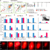
Increased brain activation during functional recovery. A, Brain areas activated during the precision grip task before the lesion were quantified in terms of increased rCBF by using H215O-PET. The rCBF during the control task was subtracted from that during the precision grip task. Brain areas with significantly increased rCBF (p < 0.05, corrected for multiple comparisons in two monkeys) are superimposed on a macaque monkey brain MRI template. The significance level is given in terms of a Z score represented on a color scale. Top row, Views from both hemispheres contralateral and ipsilateral to the hand used in the precision grip task. Bottom row, Coronal sections at the level of lines shown in the inset. AIP, Anterior intraparietal cortex; Cb, cerebellum; PMd, dorsal premotor cortex. B, C, Activation during early (B) and late (C) postrecovery periods (rCBF during the precision grip task − rCBF during the control task) was compared with that during the prelesion stage. Blue areas represent histologically confirmed lesions. B, C, Double arrowheads in the top row indicate the same subregions of the PMv; these subregions showed increased activation during the early but not late postrecovery periods.
Figure 3.
Figure 3.
A–C, Activation before the lesion and during the early and late postrecovery periods in the lesioned hand area of M1 (A), ipsilesional PMv (B), and contralesional PMv (C). Error bars indicate SD. D–F, The ROIs in the hand area of M1 (D), ipsilesional PMv (E), and contralesional PMv (F) used to calculate the bar graphs in A–C. ROIs of ipsilesional and contralesional PMv were defined as areas in which significantly increased activity was observed in the late postrecovery period (Z > 2.3). The cluster volumes were 45.1 mm3 in the ROI of M1 and 25.3 mm3 in the ROIs of ipsilesional and contralesional PMv, respectively.
Figure 4.
Figure 4.
PPI analysis revealed increased functional connectivity within M1 during functional recovery. A, B, During recovery, there was a change in the functional correlation between activity in the lesion area and activity in the perilesional M1 (arrowheads). Areas in which activation changed (p < 0.01 uncorrected, Z > 2.3) are superimposed on a 3D reconstruction of a macaque monkey brain MRI template. The brown region represents the seed region, which includes the lesion area in M1. Right panels, PPI effects around the central sulcus from a view perpendicular to the cortical surface, as shown in E. White dotted lines indicate the distance between the center of the lesion area and the peak voxel in the rostromedial M1 (early postrecovery, 3.6 mm [A]; late postrecovery, 3.8 mm [B]). The significance level is given in terms of a Z score represented on a color scale. C, D, Graphs show representative changes in interactions between activity in the lesioned area and activity in the perilesional M1 during the precision grip task in the early (C) and late (D) postrecovery periods. Blue triangles and blue regression lines represent prelesion data. Yellow crosses and red regression lines represent data during the postrecovery periods.
Figure 5.
Figure 5.
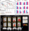
Effects of inactivating the PMv and M1 of the ipsilesional hemisphere on hand movements. A, The topographic motor representations of PMv and M1 derived before lesion, from a view perpendicular to the cortical surface. Movements elicited at the threshold of ICMS are indicated by the symbols. Some electrode penetration sites showed no response to ICMS at current strengths up to either 50 μA for M1 or 100 μA for PMv. Gray and brown areas represent areas of histologically confirmed lesions. Colored stars represent sites of muscimol injection. Arrows indicate rostral–caudal and medial–lateral orientations. R, Rostral; M, medial; asl, lower arcuate sulcus; asu, upper arcuate sulcus; cs, central sulcus. B–D, The effects of inactivating PMv, in which ICMS elicited movements of hand digits (PMv-h, B), the PMv area rostrolateral to PMv-h (PMv-rl, C), or the hand digit area of M1 (M1-h, D). The success rates of precision grip in two monkeys before the M1 lesion and during the early and late postrecovery periods after the lesion are shown. Muscimol injections into both the ipsilesional PMv-h and PMv-rl in the early postrecovery period resulted in a significant deficit in performing the precision grip in both monkeys. *p < 0.01 (Fisher's exact test). **p < 0.001 (Fisher's exact test). ***p < 0.0001 (Fisher's exact test). In the late postrecovery period, muscimol injection into either of the areas (Monkey [Mk]-Ja, PMv-h; Mk-Ki, PMv-rl) resulted in a deficit of the precision grip. E, The effects of inactivating the ipsilesional M1-h on the rate of use of an alternate grip (i.e., types of grasping other than precision grip). Inactivating the primary motor cortex (M1-h) during the late postrecovery period resulted in the impairment of movements that depend heavily on distal digits, such as the precision grip, whereas proximal movements remained intact. Muscimol injection into the ipsilesional M1-h during the late postrecovery period impaired precision grip but, in contrast to that before lesion, did not impair whole-hand grip in either monkey. Consequently, the monkeys retrieved the object by using an alternate grip, in which the object was held between the thumb and around the proximal joint of the index finger. Inset photograph, An example of the alternate grip in Mk-Ki. F, Serial coronal sections of M1 spaced by 0.8 mm showing the extent of diffusion of 3.0 μl fluorescent muscimol. Arrowheads and double arrowheads indicate the locations of the cortical surface and the boundaries between the gray and white matter, respectively. GM, Gray matter; WM, white matter. Scale bars: A, 2 mm; F, 1 mm.
Figure 6.
Figure 6.
Effects of inactivating the PMv of the contralesional hemisphere on the performance of precision grip. A, Topographic motor representation of the PMv and M1 of the contralesional hemisphere derived before lesion. Symbols and abbreviations are the same as in Figure 5A. B, C, Effects of inactivating PMv, in which ICMS elicited movements of hand digits (PMv-h, B), or the PMv area rostrolateral to PMv-h (PMv-rl, C), on the performance of precision grip. Muscimol injections into the contralesional PMv-rl in the late postrecovery period resulted in a significant deficit in performing the precision grip in Mk-Ki. ***p < 0.0001 (Fisher's exact test). D, E, Activation during the early postrecovery period (rCBF during the precision grip task; rCBF during the control task) was compared with that during the prelesion stage in each monkey: Mk-Re (D) and Mk-Hw (E). Brain areas with significantly increased rCBF are superimposed on a lateral view of the frontal lobe. F, G, Activation during the late postrecovery period was compared with that during the prelesion stage in each monkey: Mk-Re (F) and Mk-Hw (G). Bottom row, Coronal sections during the late postrecovery period at the level of the dotted lines. During the late postrecovery period, increased activation of the contralesional PMv was observed in Mk-Hw (double arrowhead) but not in Mk-Re. Significance level is given in terms of a Z score represented on a color scale.

Similar articles

Cited by

References

    1. Arikan R, Blake NM, Erinjeri JP, Woolsey TA, Giraud L, Highstein SM. A method to measure the effective spread of focally injected muscimol into the central nervous system with electrophysiology and light microscopy. J Neurosci Methods. 2002;118:51–57. doi: 10.1016/S0165-0270(02)00143-7. - DOI - PubMed
    1. Courtine G, Bunge MB, Fawcett JW, Grossman RG, Kaas JH, Lemon R, Maier I, Martin J, Nudo RJ, Ramon-Cueto A, Rouiller EM, Schnell L, Wannier T, Schwab ME, Edgerton VR. Can experiments in nonhuman primates expedite the translation of treatments for spinal cord injury in humans? Nat Med. 2007;13:561–566. doi: 10.1038/nm1595. - DOI - PMC - PubMed
    1. Dancause N, Barbay S, Frost SB, Plautz EJ, Chen D, Zoubina EV, Stowe AM, Nudo RJ. Extensive cortical rewiring after brain injury. J Neurosci. 2005;25:10167–10179. doi: 10.1523/JNEUROSCI.3256-05.2005. - DOI - PMC - PubMed
    1. Dum RP, Strick PL. Motor areas in the frontal lobe of the primate. Physiol Behav. 2002;77:677–682. doi: 10.1016/S0031-9384(02)00929-0. - DOI - PubMed
    1. Fabbri-Destro M, Rizzolatti G. Mirror neurons and mirror systems in monkeys and humans. Physiology (Bethesda) 2008;23:171–179. doi: 10.1152/physiol.00004.2008. - DOI - PubMed

Publication types

LinkOut - more resources